

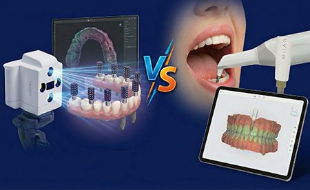


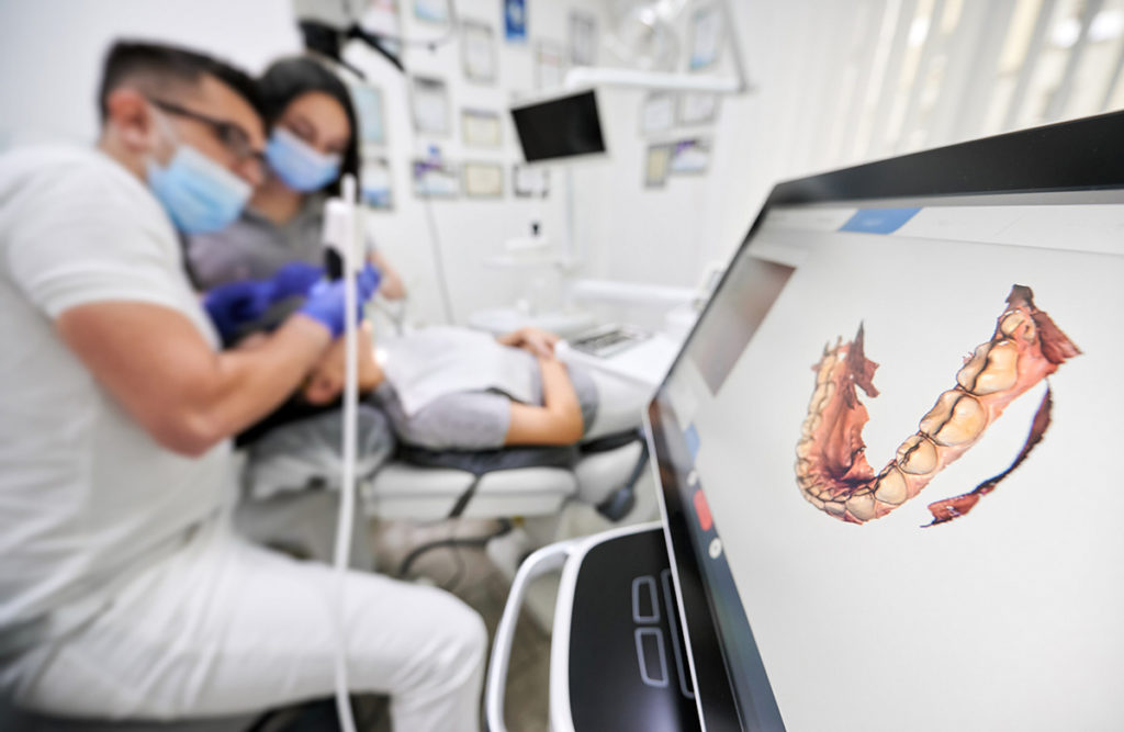
Technology has completely altered the way dentistry is performed. The introduction of digital systems has streamlined almost every workflow in the clinical setting and laboratory setting. This is especially true when considering digital impression technologies. As clinicians gain confidence in using their intraoral scanners, they naturally progress from using them to capture crown and bridge preparations to using them for more advanced processes like scanning for dental implant cases.
Benefits of Digitization of Implant Cases

Digital dentistry has created improvements to implant dentistry workflows. The ability to use an intraoral scanner (IOS) to capture the necessary data that can be used in the fabrication of precise implant restorations has created unmatched workflows. This technology has improved implant therapy in dentistry at every phase.
The benefits of using a scanner for implant cases include:
Some of these benefits are:
Steps to Ensure Accurate Digital Scans
Once an implant is fully integrated and the tissue maturation has been achieved, a digital impression scan can be done. In order to provide implant cases that offer precision and esthetics all in a timely manner, proper scanning techniques must be followed:
1. The Scan Body
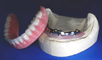
The scan body is used to determine the accurate orientation and position of the dental implants. These components are screwed into the implant fixture. With the scan bodies in place, the patient’s mouth can then be scanned with the intraoral scanner. The scan body is specific to the brand of implant that is being used. They function similarly to an impression coping used in traditional implant cases.
2. Selection of the Scan Body
There are two options when selecting scan bodies.
The first is to choose scan bodies from a major implant manufacturer. This option can usually be expected to be at a higher price point and take more time.
The second option, which has been very successful at Burbank Dental Lab, is to use scan bodies from 3rd party manufacturers. Companies like Medentika and Dess support most implant platforms. This option provides precision parts that offer a lower price point and faster turnaround times.
3. Placing the Scan Body
After the tissue is completely healed and the patient is ready for the final restoration, the scan body must be placed.
To get the best restorative outcome, the adjacent teeth should be evaluated to ensure that there are broad proximal contacts to avoid black triangles and food impaction.
The key is to achieve an ideal path of insertion for the final restoration. Any needed adjustments to the adjacent teeth should be made prior to scanning the area.
4. Checking for Accuracy
It is imperative that the scan body be completely seated. Once the scan body is placed, it should be checked with a radiograph to ensure proper placement. This scan will result in a final abutment and restoration.
Check that the scan body is placed correctly, or the final prosthesis will be inaccurate.
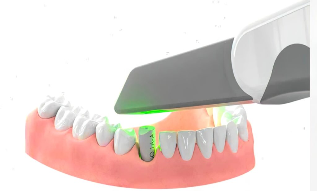
5. The Scan Process
It is necessary to scan the implant site when the scan body is in place.
However, when scanning the arch, the scan body can be removed and replaced with a provisional or healing abutment.
To achieve the best scan, do the following:
- Completely dry the soft and hard-tissue surface
- Include the scan body, adjacent dentition, contacts, occlusion, and the surrounding soft tissue in the scan.
- The scans should be done at a slow and thorough pace to ensure accuracy in the final output.
6. Taking the bite
At this point, the scan body should already be removed and replaced with either a provisional or healing abutment.
The patient should be directed to bite down to achieve maximum intercuspal positioning.
Scan and capture the bite.
7. Evaluate the scan data
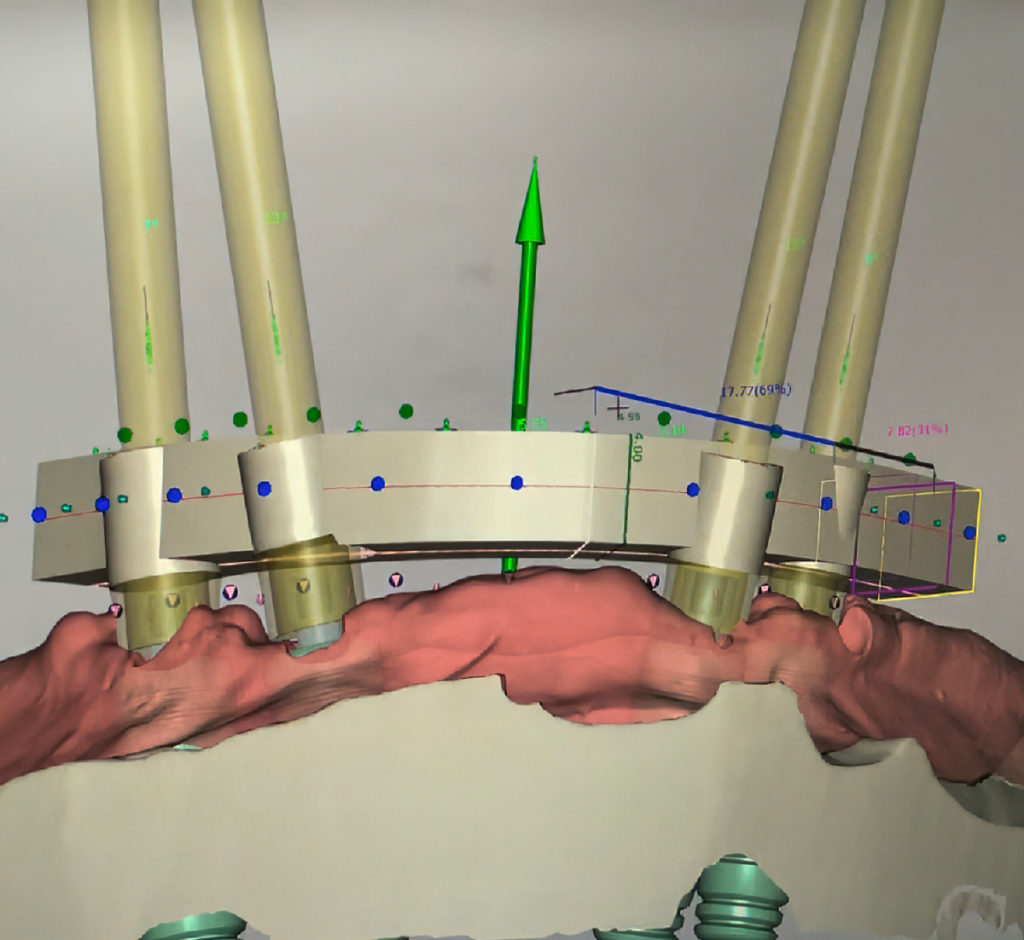
Select the scanner’s model mode when evaluating the data. This will allow for better visualization of the scan data.
Now, look for any issues with the scans, such as distortions in the scan body, missing tissue, or saliva contamination. A grainy-looking scan is usually the result of improperly drying the hard and soft tissue prior to scanning.
As with traditional impression methods, digital impressions require accuracy to allow for precision in the final restorations.
One of the great benefits of a digital workflow is the ability for the clinician to review the impression prior to sending it off to the lab. If something is off, it can simply be rescanned and evaluated. This greatly improves patient satisfaction, turnaround time, and accuracy.
Call or chat with one of Burbank Dental Lab’s implant team members for help with your next implant case.