Digital Technology: Improvements in Implant Dentistry

mm Written by Andrew Sedler

Dental implants have proven to be a highly successful treatment option for cases where edentulism is present. One of the most challenging restorative procedures facing both clinicians and laboratories is restoring a single anterior tooth where an implant is involved. Using dental implants is one of the most conservative options, as the alternative would require fabricating a three-unit bridge.

The key area to the success of these types of cases lies in the fundamentals of the fabrication process itself. Digital Dentistry has helped create exceptional workflows for single unit implant cases.

Digital Impressions
Digital Methods
Figure 1

implant information needs to be transferred in a highly accurate manner to achieve precision in the final prosthesis. Digital impressions offer a sound way to transfer implant data to the laboratory. Intraoral scanners have been found to provide a high level of precision in the transfer of precise oral data. In addition, intraoral scanners offer the following benefits:

  • Patients do not experience the gagging and bad taste often associated with conventional impression methods.
  • Accuracy is one of the key areas to ensure implant survival and provide optimal esthetic outcomes. Digital methods (Fig. 1, see above) streamline the process, reducing the opportunity for error by reducing steps and the possibility of the instability of dental materials found in traditional impression methods.
  • Impression material and trays are eliminated, reducing overhead.
  • Digital impressions are a faster way to impress, thereby saving valuable chair time.
  • Elimination of having to ship impressions to the lab shaves days off the turnaround time,
  • Remakes are reduced since the data can be reviewed while the patient is still in the chair. The data can be shared with the lab and discussed in real-time. Problems are found before fabrication, saving time and money.
Scan Bodies
Scan Body

To achieve this, scannable impression copings or scan bodies are used. Most implant manufacturers offer a specific scan body (Fig. 2, see above) that provides the position of the implant, including height and angle. This is done by screwing the scan body into the implant, creating a virtual impression coping. The area is then scanned with an intraoral scanner. The opposing and bite information is also recorded, and this information is used to create a digital model. As with any impression-taking method, the accuracy of the data is critical to the accuracy of the final result. For best results in creating a digital implant scan, we recommend the following:

  1. Utilize Burbank Dental Lab's implant specialists. They will help determine restorative options, pricing, scan body types, and any other issues regarding the case before the scan.
  2. To accurately digitize the case, make sure to provide a scan of the scan body, opposing, soft tissue, and bite.
  3. Over time, scan bodies can show signs of wear. Evaluate scan bodies to ensure they are damage free to ensure accuracy in the scan.
Digital Implant Scan Workflow

Once the implant has integrated and the tissue is healed, the restorative process can begin. This step requires the patient to be scanned with an intraoral scanner to gather the implant and oral cavity digital data. Here the scan body is placed into the implant. The proper placement can be determined with a radiograph.

Once proper seating of the scannable coping is determined, the case can be scanned. Once the scan body, opposing, and bite are recorded, a 3-dimensional model is created on the screen. At this point, an evaluation is done to ensure all of the critical information has been captured, including the scan body, the tissue, the opposing, the bites, and the adjacent teeth.

After the scan has been completed and evaluated, detailed instructions must be sent to Burbank Dental Lab. These instructions should include the type of abutment desired, the shade, the final restoration material, whether the case is cement or screw-retained, and any other information necessary to provide the desired results. A Burbank Dental Lab implant specialist is available to help with any issues regarding selecting the restorative options that are optimal for a case.

Success Guides of Burbank Dental Lab
Restoration Fabrication

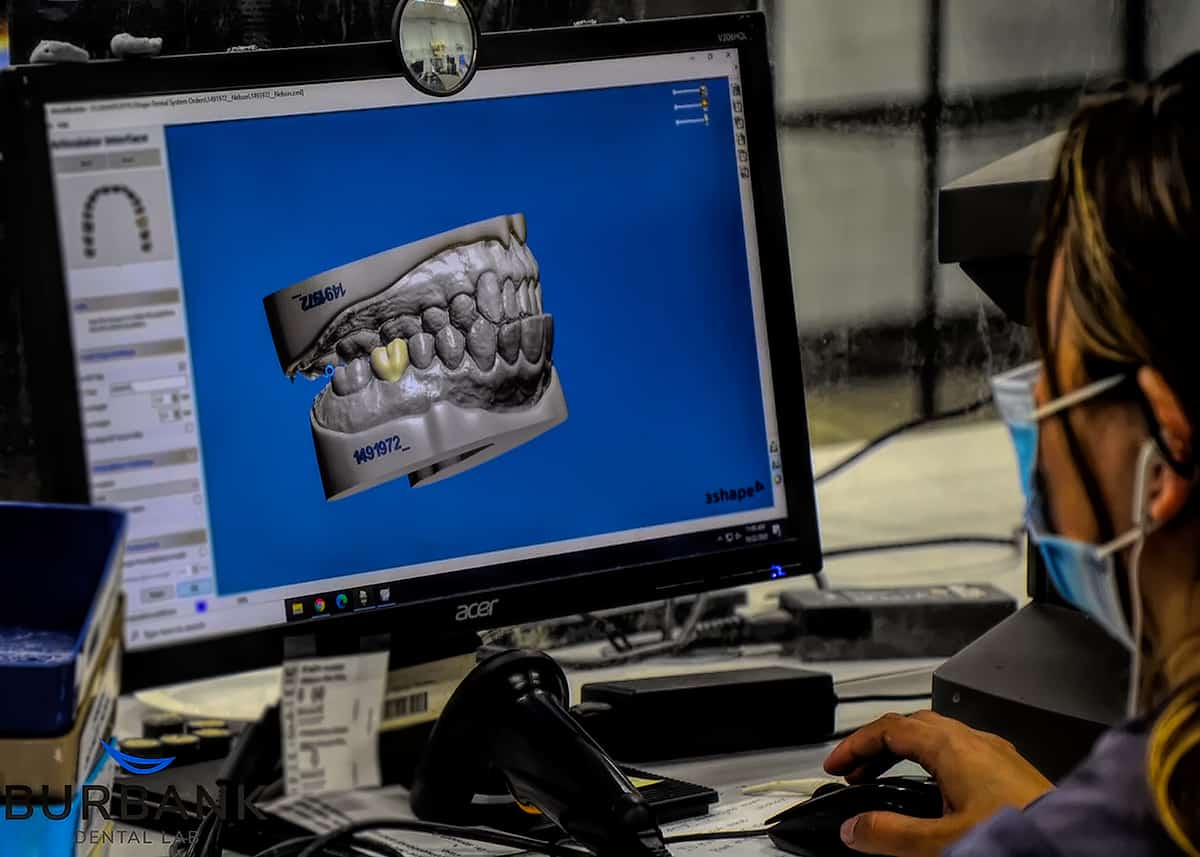
Burbank Dental Lab then utilizes this detailed information to begin designing the restoration. The data is uploaded to advanced design software, and the occlusion/bite, restorative position/space, and accuracy are evaluated. If there are any issues, the clinician can be alerted before the restoration is created.

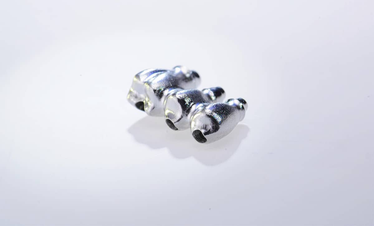
Once the evaluation is complete, the abutment is designed along with either a full-coverage restoration or coping. The appropriate thickness, height, and access hole (screw-retained) (Figure 3, see image below) placement are determined.

This entire workup is completed digitally.

screw-retained
Figure 3

The data is then sent and queued to begin printing a working model, milling the abutment, and milling the appropriate restoration. Along with all of the steps mentioned above, the equipment used to both design and mill the restorations is crucial to these types of cases' accuracy and success.

Burbank Dental Lab is dedicated to investing in the most advanced technology to ensure that each restoration produced is of the highest degree of accuracy. The equipment a laboratory uses to mill out restorations matters in terms of the precision of the restorations.

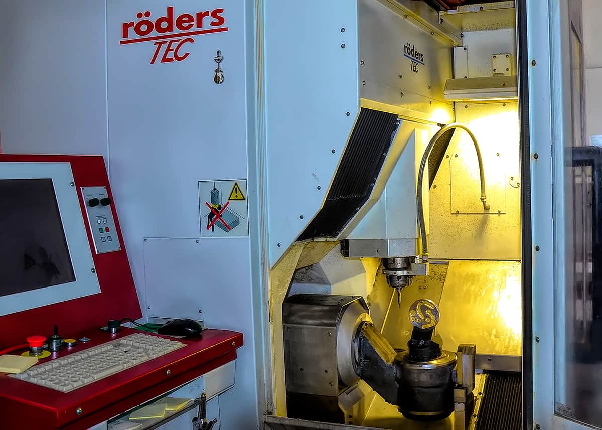
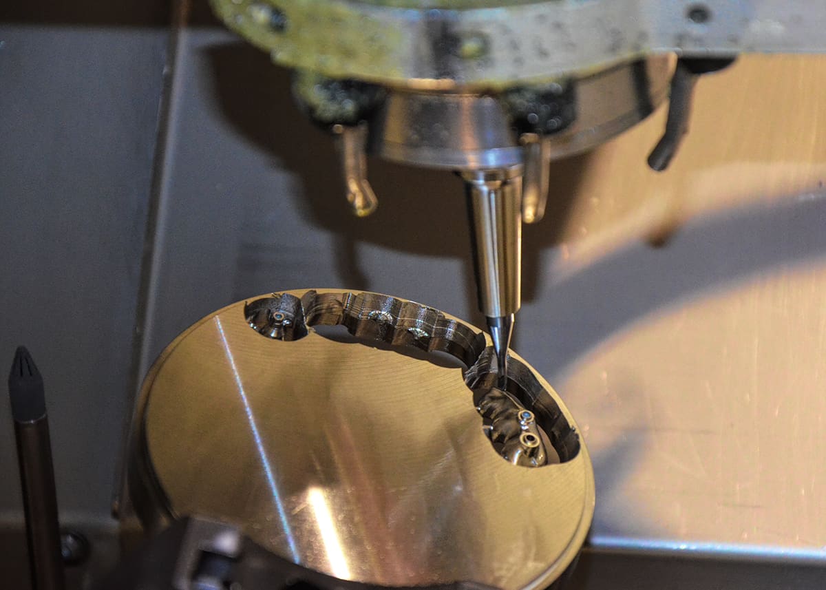
At Burbank Dental Lab, our outputs are done using CNC (computer numerical control) machinery with up to 5-Axis, offering the highest precision. Our milling machines take the data from our designers (CAD) and convert them into numbers (Figure 4, see image below). These numbers are then used to control the movement of the milling machine.

The more sophisticated the process, the more precise the cutting and shaping of the final product. The precision of restoration is essential for the integrity of margins and contacts. But it also is crucial to the customization of implant abutments.

Milling Machines
Figure 4

Digital dentistry is moving exponentially in terms of innovation. While these innovations have disrupted the dental industry, they have created new pathways for delivering improved accuracy. Unquestionably, implant dentistry has improved dramatically thanks to digital dentistry workflows. The technology provides for improved impression techniques, communication, and design. As with any technology, there are several options available. At Burbank Dental Lab, we are heavily invested in research and development to ensure that we incorporate the most advanced options the industry has to offer.

Did you like this article?

Let us know by giving us a clap!

2+

Top 10 Most Clapped Articles

Resource Center

Products

Slide 1

Fabrication
of the future
is here!

Burbank Dental Lab has three new state-of-the-art Carbon M2 printers. We are very excited about the options that these cutting-edge printers will allow us to offer our dental clients. Here are some of the advantages that these printers will begin to deliver to you and your dental practice.

Our New
State-Of-The-Art
3d Printers

Carbon offers a highly dependable 3D manufacturing solution for many dental applications with its breakthrough Digital Light Synthesis™ technology, enabled by a wide range of dental materials.