Perfecting the Single-Unit Case with Digital Dentistry

Perfecting the Single-Unit Case with Digital Dentistry

ADVANCED TECHNOLOGIES LEAD TO IMPROVED DIAGNOSIS

Written by Andrew Sedler, COO

Written by Andrew Sedler, COO

Written by Andrew Sedler, COO

At Burbank Dental Lab, we believe that advanced technologies lead to improved diagnosis, case planning, and, ultimately, delivery of the final restorations.

Digital impressions have greatly improved the entire implant treatment digital workflow. Digitization has made the process more efficient and has enhanced the entire experience for the patient.

The use of technology is becoming the standard of care by many clinicians, helping to advance the processes and lead to more innovative solutions that ultimately improve case outcomes.

Implant restorations are one of the fastest-growing areas in dentistry. It is overwhelmingly a superior treatment option for single tooth replacement.

Other treatment options, like three-unit bridges, involve the restoration of healthy teeth next to the edentulous area. These types of fixed prostheses can cause periodontal or endodontic complications of the supporting abutments, creating unnecessary trauma.

Implants provide favorable outcomes that include esthetics, function, and help in preserving the surrounding bone. Implant cases require proper planning and case selections to achieve success.

Case Planning

Evaluation then begins with determining the viability of implant placement.

Bone density is a key factor and can be determined by radiograph evaluation and standard measurements. Evaluation is done of the alveolar ridge to determine if adequate length, width, and depth are present to insert an implant.

Also, crown height space should be evaluated by the clinician. To do this, one should measure from the crest of the alveolar ridge to the occlusal plane. Adequate space is needed between the ridge and opposing dentition to achieve proper esthetics, function, and strength. In addition, evaluations of the following should be done:

  • Periodontal evaluation to assess both the hard and soft tissue
  • Evaluation of adjacent dentition inter-root space
  • Occlusal analysis to determine the proper occlusal scheme to help ensure the implant restoration's longevity during normal mastication forces.
Using A Diagnostic Wax-Up

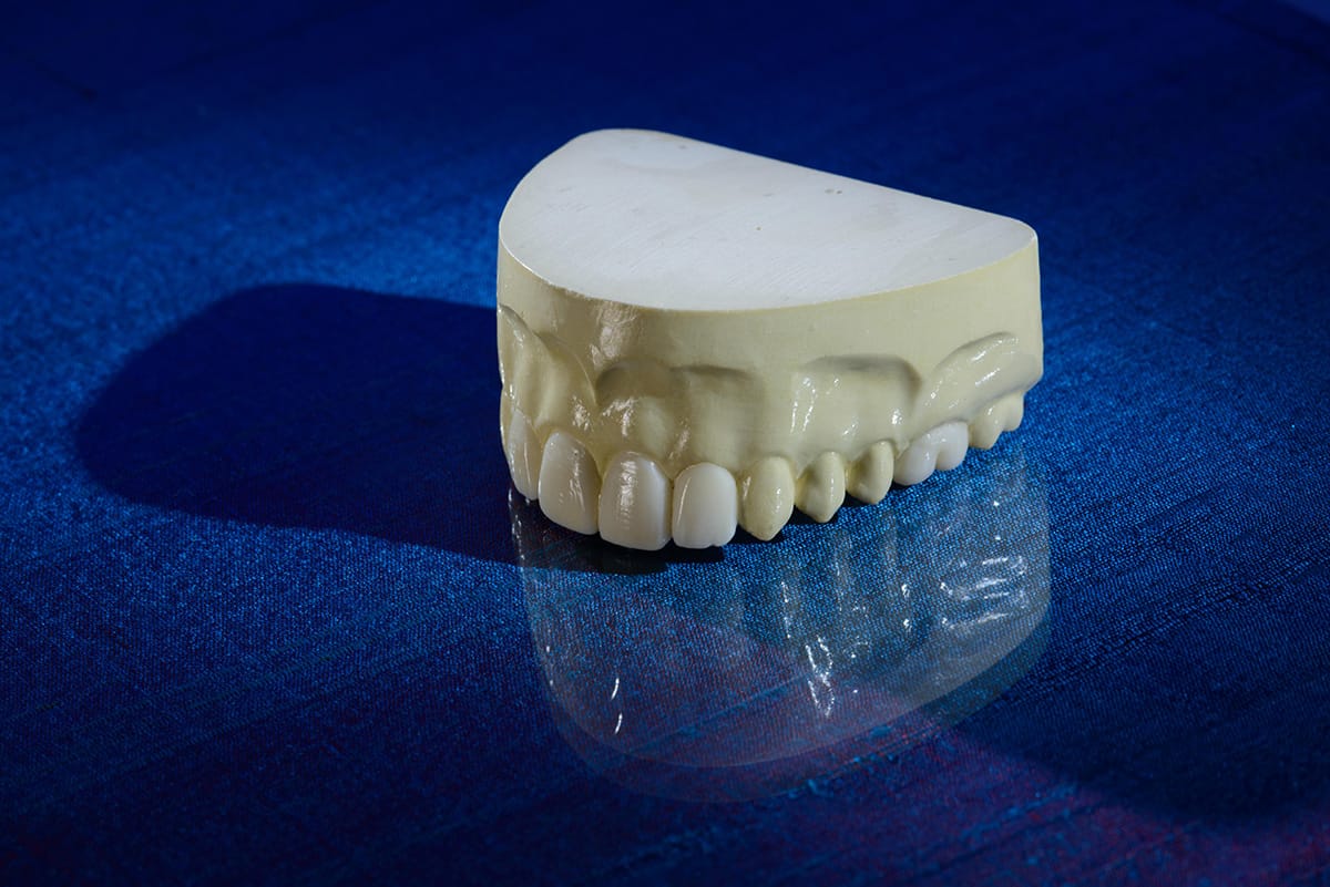
In some cases, it may be necessary to complete a diagnostic wax-up.

This wax-up can help to determine what is possible and how to achieve optimal results as they relate to the tissue. From this, a model-based surgical guide is fabricated. This can help in determining the correct angulation and placement.

These guides are fabricated out of the following materials:

  • Metal cylinders
  • Gutta-Percha
  • Radio opaque acrylic
Digital Impressions
CBCT Cone Beam Computerized Tomography

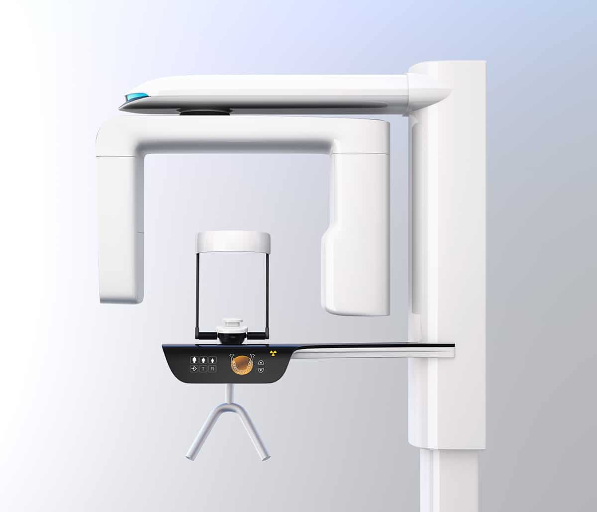
A cone-beam computerized tomography (CBCT) scan can help with the proper placement of the implant.

The patient receives both a CBCT scan and an intraoral digital scan. The data from the CBCT scan and the data from the intraoral scan are merged.

Next, a surgical guide is designed from the merged file. The file is then sent to Burbank Dental Lab, where a surgical guide is fabricated.

The Surgical Guides at Burbank Dental Lab provide the following:

  • The guide is checked on a solid model to ensure proper fit.
  • Precision guide rings are used in each appliance unique to each surgical system.
  • Only FDA approved resin is used
  • Quick turnaround time

The surgical guide is then used to properly place the implant. After osseointegration has occurred, the restorative process begins.

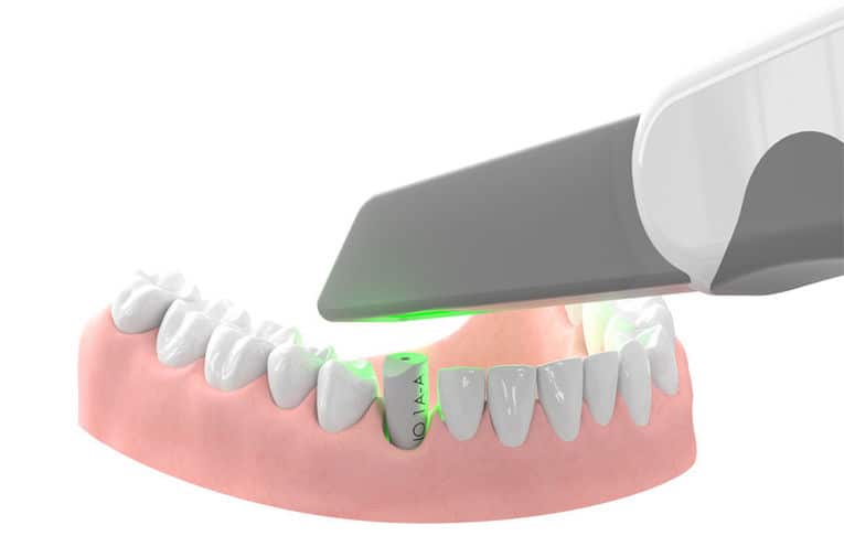
The use of intraoral scanners to take digital impressions offers excellent accuracy for single-unit implant restorations.

Once a scan is done, it provides an immediate connection with the laboratory. The file can be shared and discussed.

Digital impressions eliminate the need for traditional impression materials, reducing overhead. Due to the efficiency and accuracy of intraoral scanners, valuable chair-time is significantly reduced. It has been found that clinical adjustment times are shorter when a digital workflow is used over a conventional one. Also, digital impression taking is overwhelmingly preferred by patients over the use of chemical impression methods.

Scan Bodies for Digital Scans
Scan Bodies - Burbank Dental Lab

Scan Bodies are a vital part of ensuring an accurate digital scan. Most major implant manufacturers have developed a scan body that relates the brand, height, angle, and overall position of the implant. This information is then transferred to a digital model. In addition to this, the opposing and bite are captured through the digital impression process.

For best results, we recommend the following:

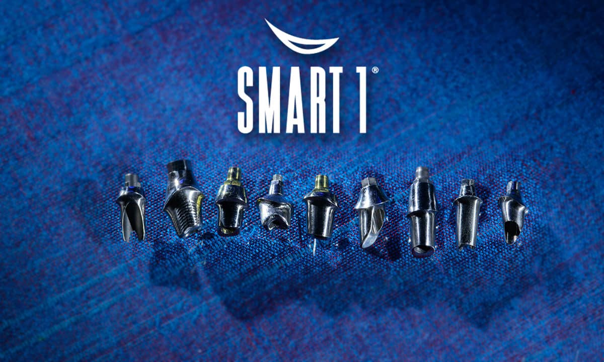
  1. Call Burbank Dental Lab to speak to one of our implant specialists regarding the Scan Body types (see image above). This step can help provide information related to restoration choices, pricing, and turnaround times.
  2. To ensure accuracy in the scan, please provide the following scans:
    - Antagonist
    - Scan Body
    - Soft tissue scan
  3. Make sure the scan body being used is free from damage. It must be in good condition to accurately relay vital implant information.

After all of the data has been scanned and sent to the laboratory, the abutment is designed.  Smart1 implant abutments (see image below) from Burbank Dental Lab are proprietary abutments used in either cement-retained or screw-retained cases.

These abutments are designed digitally and are considered custom abutments. This customization is used to create perfect tissue contouring by carefully designing the emergence profile. They are either fabricated out of titanium or zirconia with a titanium base. The material choice is dependent on case goals.

Creating the Perfect Final Restoration

The final restoration is then designed and milled. One great option for restoring implant cases is zirconia.

Zirconia is milled using computer-aided design and computer-aided manufacturing. When esthetics is a critical consideration, zirconia coping is milled, and then porcelain can be layered. This is great for matching dentition.

Digital design and manufacturing are becoming more accepted every day. This technology helps to simplify processes while increasing accuracy, lowering overhead, and improving the patient experience.

Clinical adjustments have become faster as a result of improved accuracies than when conventional methods are used. The implant team at Burbank Dental Lab is available to help guide you through your next implant case.

Did you like this article?

Let us know by giving us a clap!

1+

Top 10 Most Clapped Articles

Resource Center

Products

Slide 1

Fabrication
of the future
is here!

Burbank Dental Lab has three new state-of-the-art Carbon M2 printers. We are very excited about the options that these cutting-edge printers will allow us to offer our dental clients. Here are some of the advantages that these printers will begin to deliver to you and your dental practice.

Our New
State-Of-The-Art
3d Printers

Carbon offers a highly dependable 3D manufacturing solution for many dental applications with its breakthrough Digital Light Synthesis™ technology, enabled by a wide range of dental materials.