


The best digital practices
aren’t choosing sides
— they’re using both.
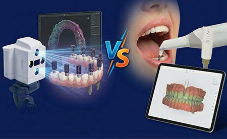
Photogrammetry vs. IO Scan Body
Modern dentistry is rapidly embracing digital workflows, especially in dental implantology, where precision is critical to case success. Two methods for capturing implant positions digitally are Photogrammetry and the IO scan body technique.
Photogrammetry uses photographic images to pinpoint implant locations with high accuracy, while the IO scan body method employs intraoral scanners and specialized scan bodies to digitally record implant positions. Both approaches aim to replace or enhance traditional impressions, offering dentists improved accuracy, efficiency, and patient comfort.
What is Photogrammetry in dentistry?
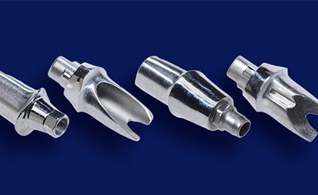
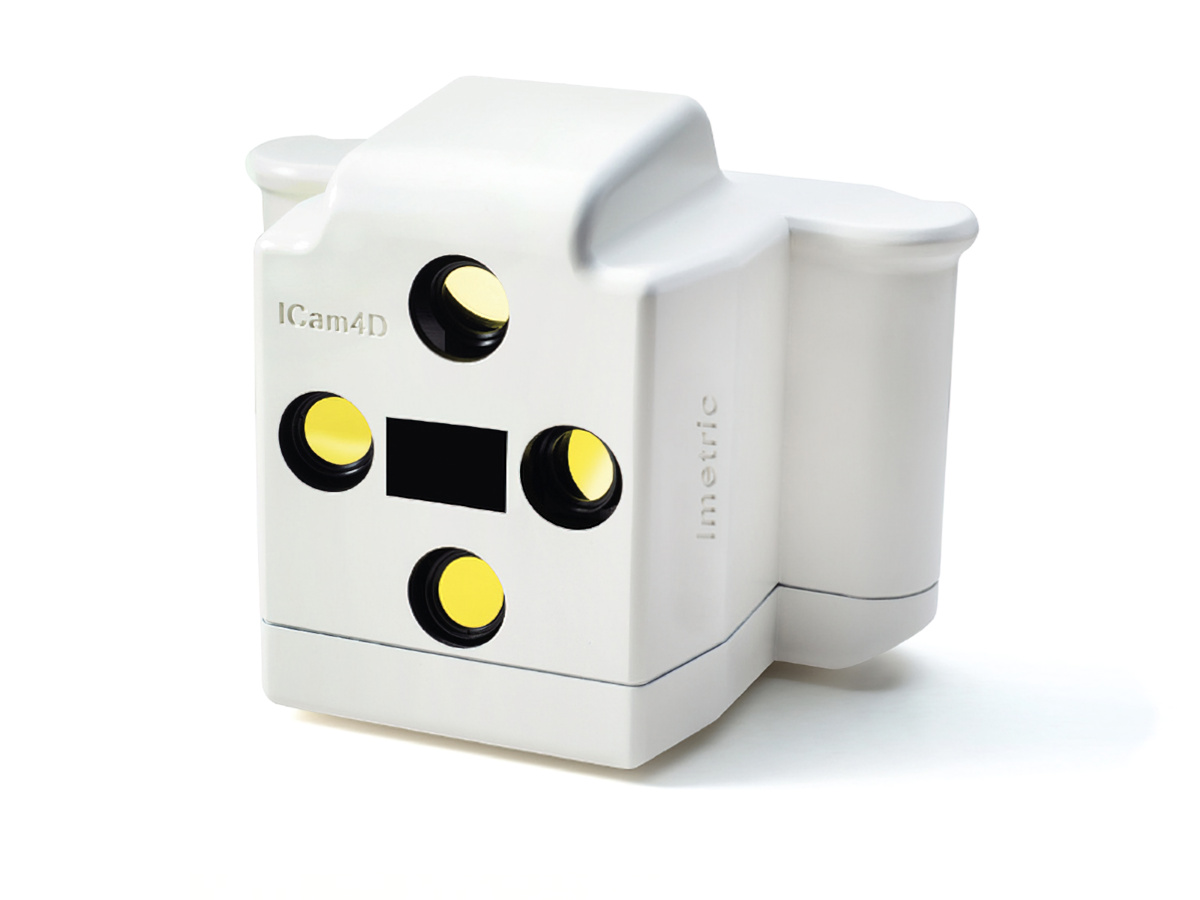
Photogrammetry in dentistry is a digital impression technique that captures the three-dimensional positions of implants using photographs. In a photogrammetry workflow, special markers or scan abutments (often called photogrammetry scan bodies) are attached to multi-unit abutments. A specialized camera then takes multiple images from different angles, and software triangulates the exact spatial coordinates of each of the positions. Through this process of stitching 2D images together, a precise 3D representation of the implant positions is created.
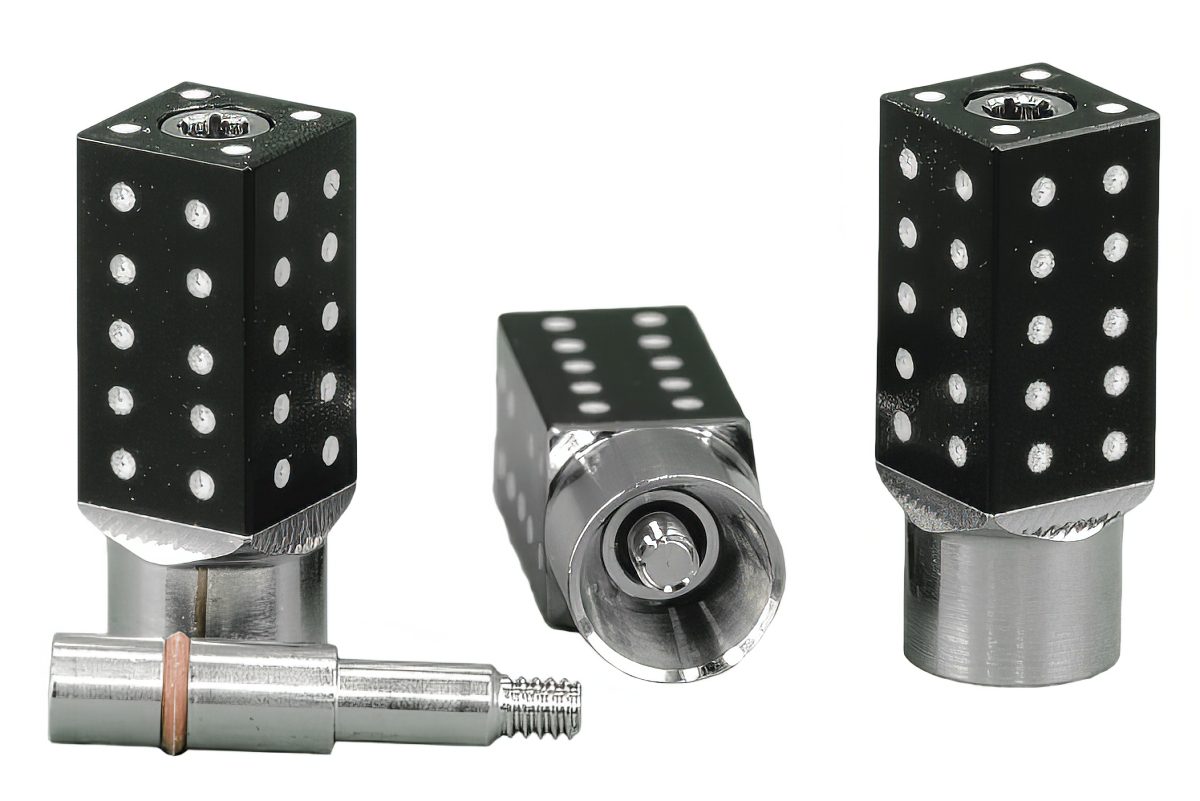
Photogrammetry essentially treats each implant’s location as a “target” to be measured. Unlike conventional intraoral scanning, the photogrammetric capture is done extraorally – the camera records the scan markers from outside the mouth. This extraoral capture means factors such as saliva, bleeding, or patient movement have minimal impact on accuracy. As long as the camera can see the white dots on the markers clearly, it will record the positions.
Saliva, bleeding, and patient movement? Photogrammetry doesn’t care.


Dr. Gent Memetic, DMD
“I’ve been using Burbank Dental Lab for all my dental cases, and they’ve been fantastic to work with. The quality of their work is always consistent — everything fits great and looks beautiful, which makes my life a lot easier.”

Full-arch accuracy isn’t just about the technology — it’s about choosing the right technology for the case.
Benefits of Photogrammetry
What is the photogrammetry workflow?
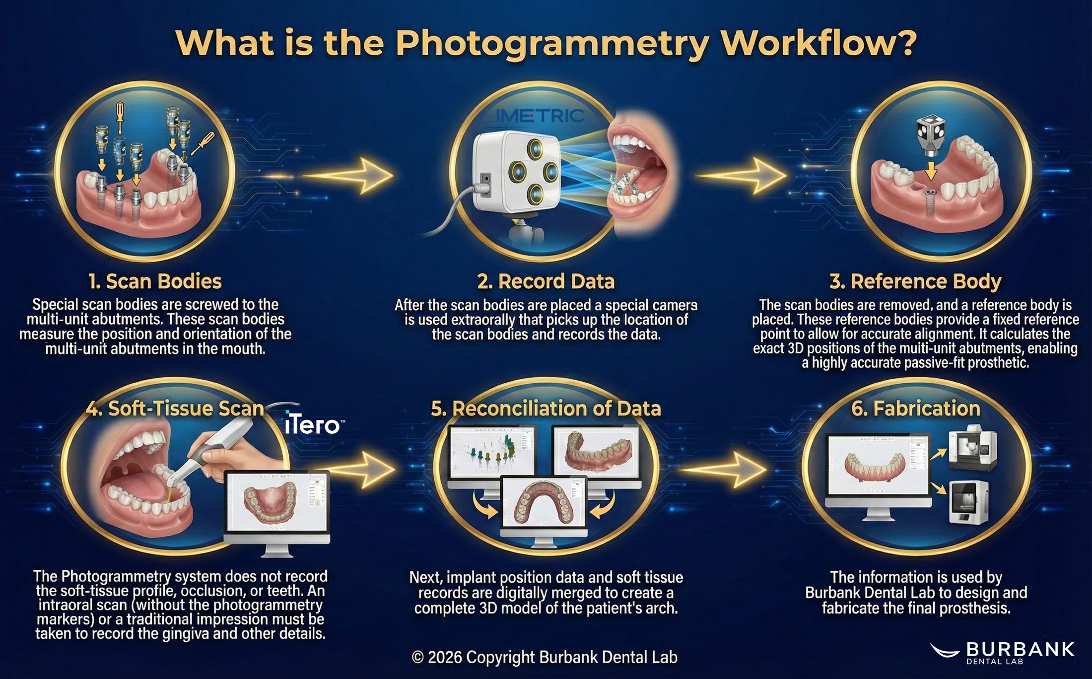
Photogrammetry focuses solely on capturing implant positions. It does not capture the surrounding soft tissue or any detailed anatomy of the gums and teeth. Therefore, the process is as follows:
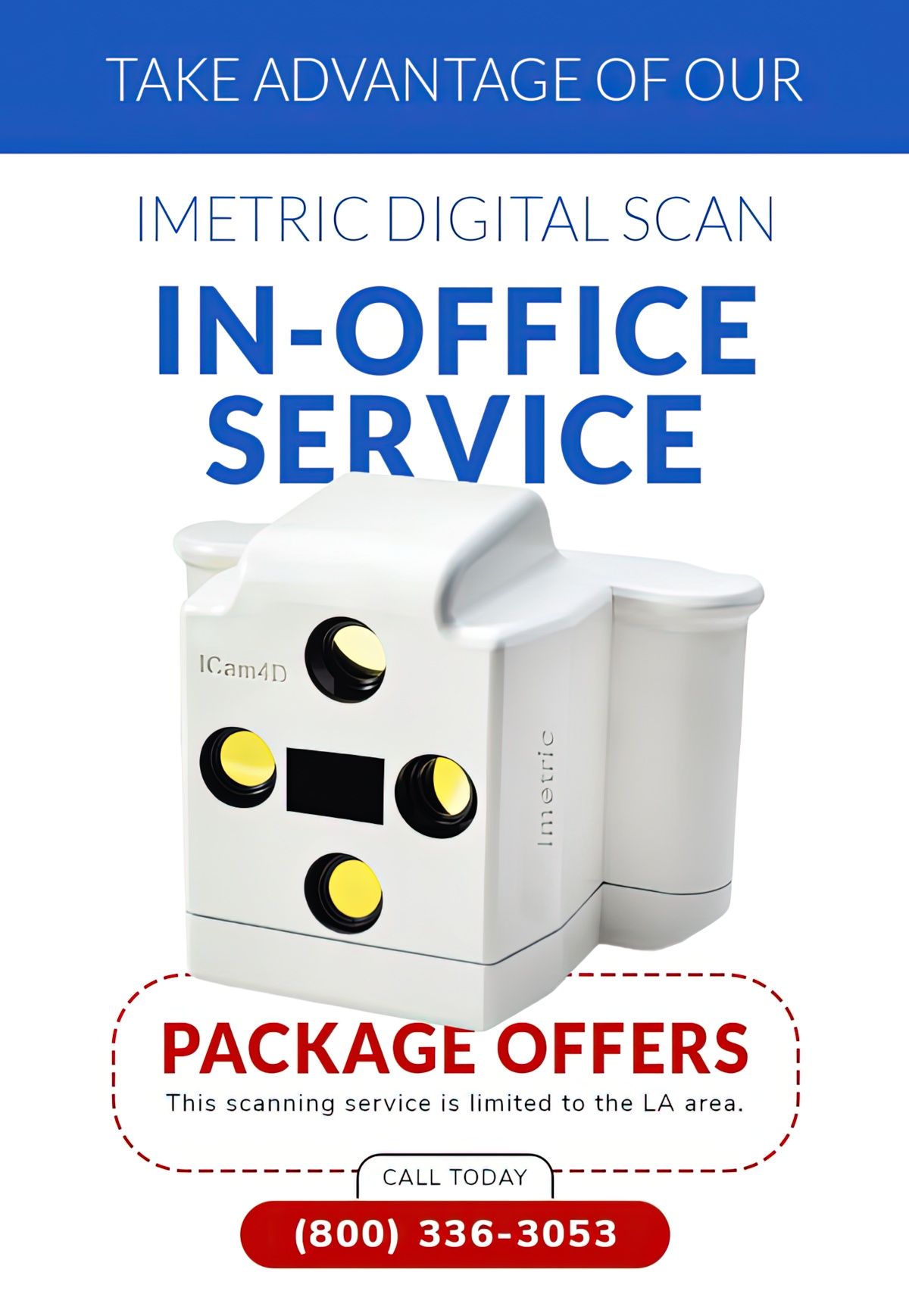
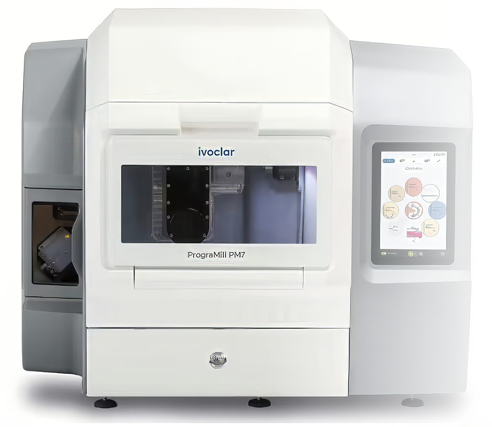
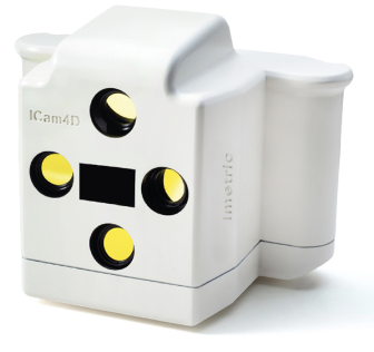
Current prominent photogrammetry systems on the market include iCam4D (iMetric) and the PIC Camera (PIC Dental), among others.
You don’t need to invest in a photogrammetry system —
Burbank Dental Lab
already has one!
One method captures everything in a single scan — the other delivers unmatched precision in two steps.
FREE TO DOWNLOAD – SUCCESS GUIDES
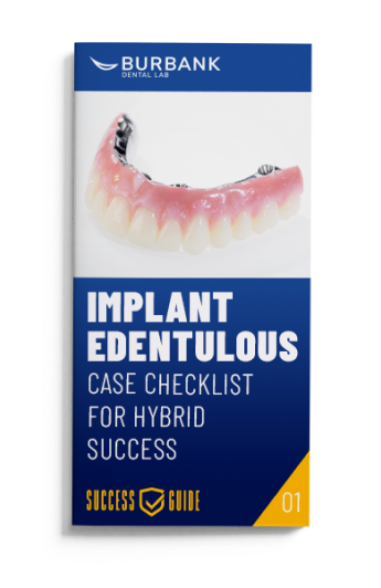

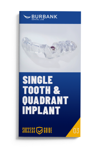
DOWNLOAD A GUIDE
What is an IO Scan Body (intraoral scanning) technique?
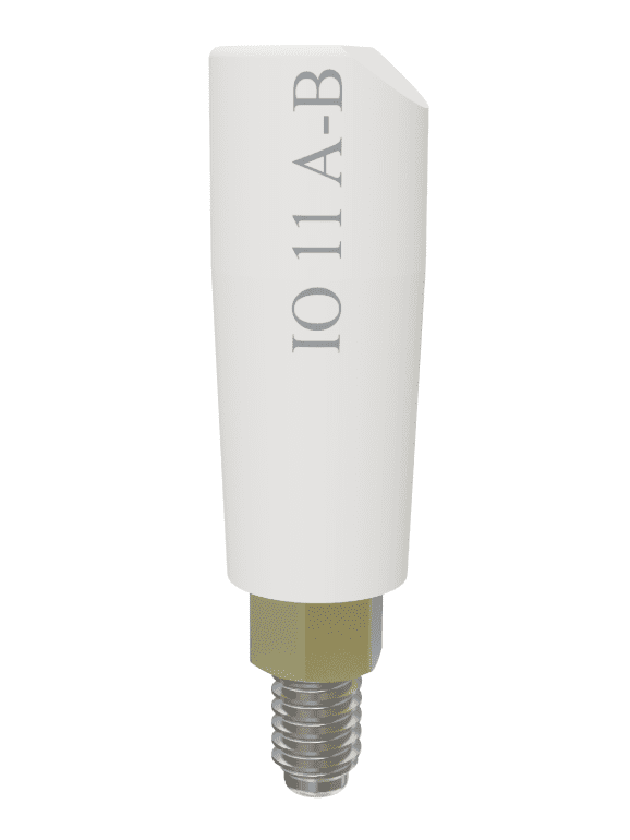
IO scan bodies, such as the Elos Accurate® Scan Body, are used with intraoral scanners to capture digital implant impressions. A scan body (also called an intraoral scan body) is a small, precisely manufactured component that attaches to a dental implant. It serves as a reference object for the scanner because the scan body has a specific shape with known dimensions and features; the scanning software can recognize it and pinpoint the implant’s location and orientation in the digital model. They are screwed or snapped into the implant in place of a healing abutment or temporary crown during the scan.
For single implants, your intraoral scanner already has you covered.
Benefits of IO Scan Body technique
The following are the benefits of the IO scan body technique:
The best full-arch solution isn’t always the most fixed one—it’s the most appropriate one.
What is the IO Scan Body workflow?
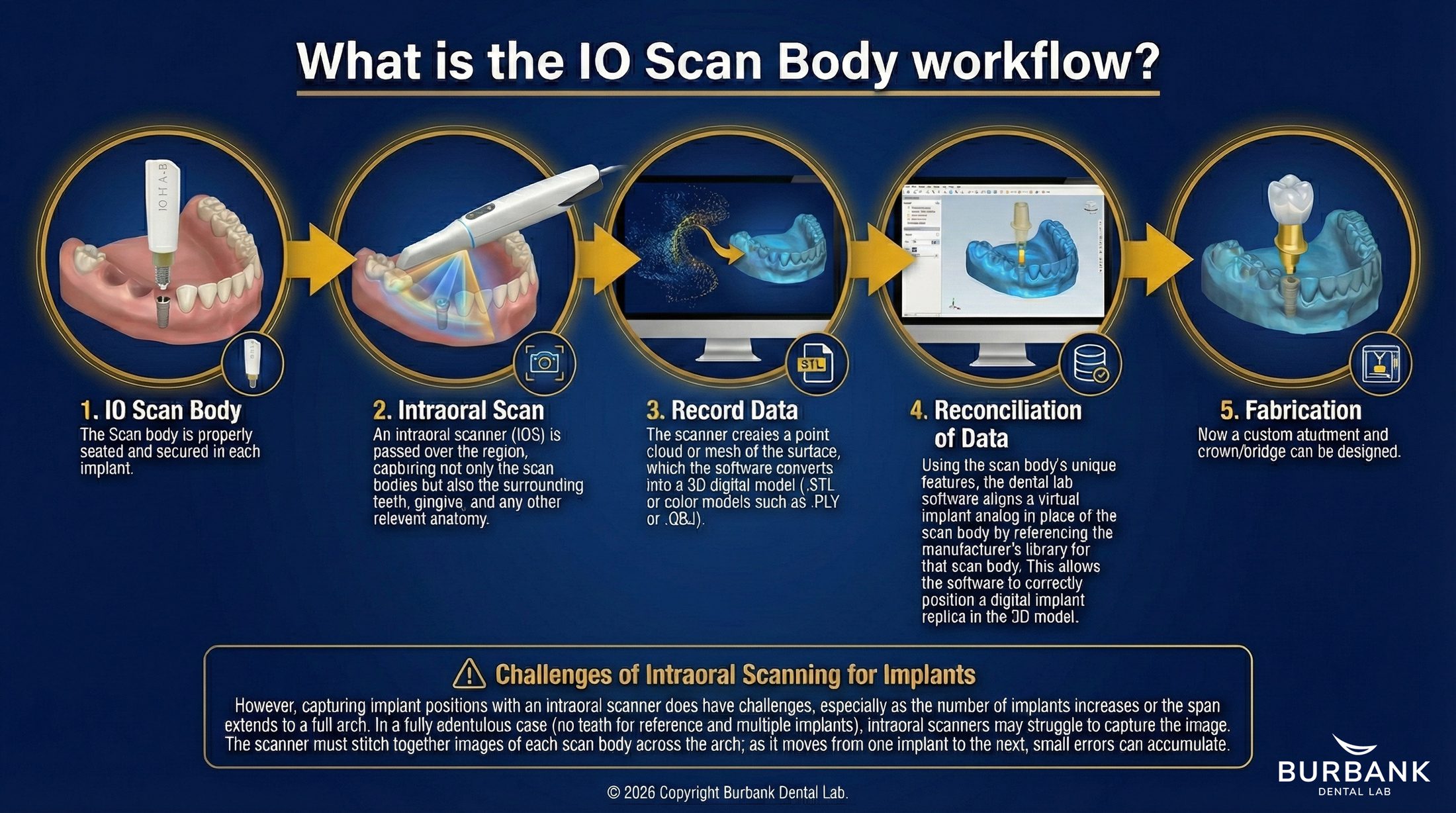
However, capturing implant positions with an intraoral scanner does have challenges, especially as the number of implants increases or the span extends to a full arch. In a fully edentulous case (no teeth for reference and multiple implants), intraoral scanners may struggle to capture the image. The scanner must stitch together images of each scan body across the arch; as it moves from one implant to the next, small errors can accumulate.
Photogrammetry vs. IO Scan Body: Key Differences
Both photogrammetry and IO scan body techniques enable digital implant impressions, but they differ significantly in methodology and ideal use cases. Below is a comparison of key aspects:
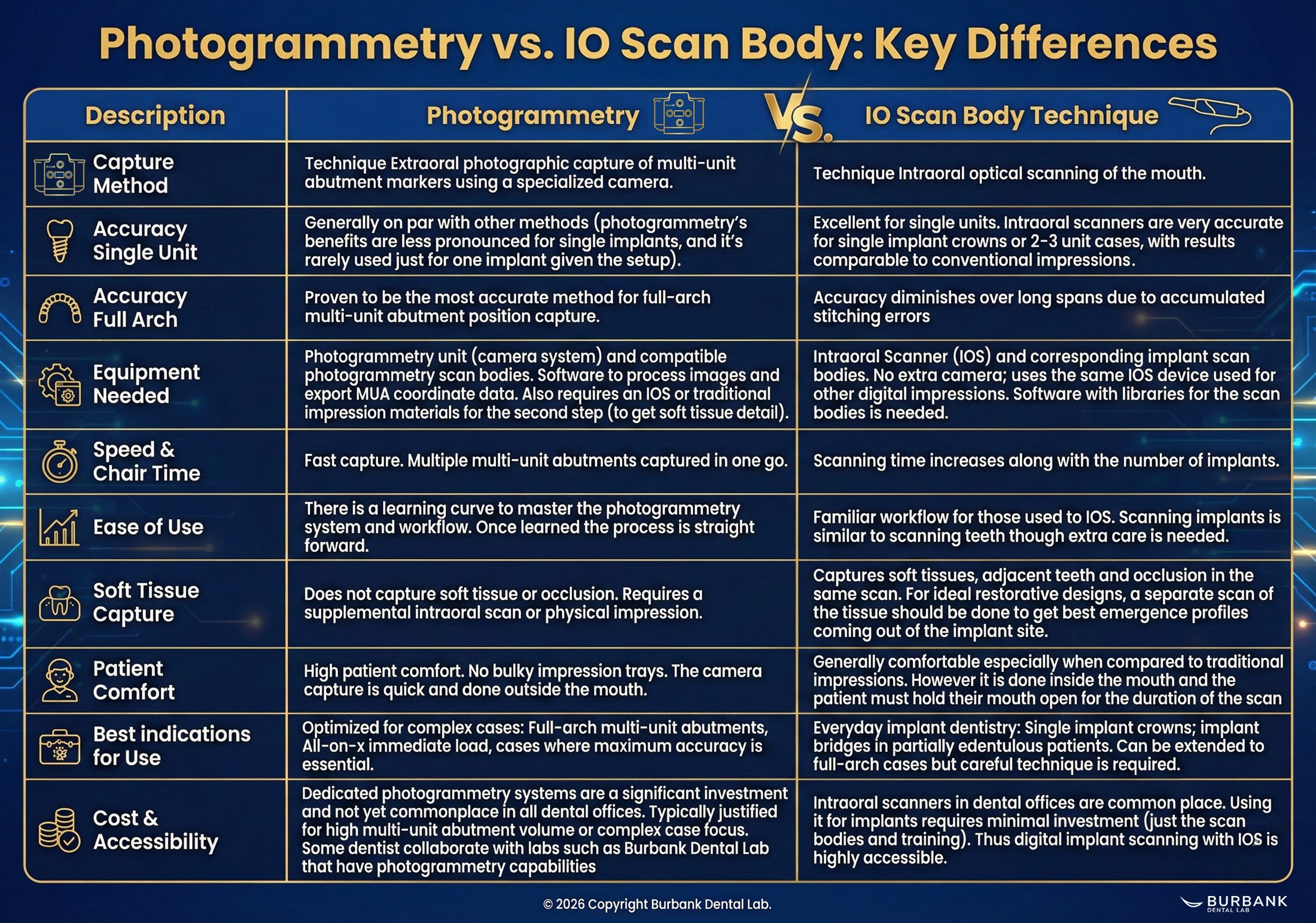
Description | Photogrammetry | IO Scan Body Technique |
Capture Method | Photogrammetry: Technique extraoral photographic capture of multi-unit abutment markers using a specialized camera. | IO Scan Body: Technique Intraoral optical scanning of the mouth. |
Accuracy Single Unit | Photogrammetry: Generally on par with other methods (photogrammetry’s benefits are less pronounced for single implants, and it’s rarely used just for one implant given the setup). | IO Scan Body: Excellent for single units. Intraoral scanners are very accurate for single implant crowns or 2–3 unit cases, with results comparable to conventional impressions. |
Accuracy Full Arch | Photogrammetry: Proven to be the most accurate method for full-arch multi-unit abutment position capture. | IO Scan Body: Accuracy diminishes over long spans due to accumulated stitching errors |
Equipment Needed | Photogrammetry: Photogrammetry unit (camera system) and compatible photogrammetry scan bodies. Software to process images and export MUA coordinate data. Also requires an IOS or traditional impression materials for the second step (to get soft tissue detail). | IO Scan Body: Intraoral Scanner (IOS) and corresponding implant scan bodies. No extra camera; uses the same IOS device used for other digital impressions. Software with libraries for the scan bodies is needed. |
Speed & Chair Time | Photogrammetry: Fast capture. Multiple multi-unit abutments captured in one go. | IO Scan Body: Scanning time increases along with the number of implants. |
Ease of Use | Photogrammetry: There is a learning curve to master the photogrammetry system and workflow. Once learned the process is straight forward. | IO Scan Body: Familiar workflow for those used to IOS. Scanning implants is similar to scanning teeth though extra care is needed. |
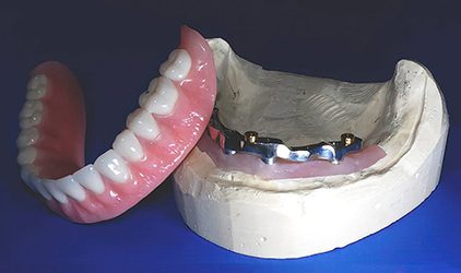
Soft Tissue Capture | Photogrammetry: Does not capture soft tissue or occlusion. Requires a supplemental intraoral scan or physical impression. | IO Scan Body: Captures soft tissues, adjacent teeth and occlusion in the same scan. For ideal restorative designs, a separate scan of the tissue should be done to get best emergence profiles coming out of the implant site. |
Patient Comfort | Photogrammetry: High patient comfort. No bulky impression trays. The camera capture is quick and done outside the mouth. | IO Scan Body: Generally comfortable especially when compared to traditional impressions. However it is done inside the mouth and the patient must hold their mouth open for the duration of the scan |
Best indications for Use | Photogrammetry: Optimized for complex cases: Full-arch multi-unit abutments, All On X immediate load, cases where maximum accuracy is essential. | IO Scan Body: Everyday implant dentistry: Single implant crowns; implant bridges in partially edentulous patients. Can be extended to full-arch cases but careful technique is required. |
Cost & Accessibility | Photogrammetry: Dedicated photogrammetry systems are a significant investment and not yet commonplace in all dental offices. Typically justified for high multi-unit abutment volume or complex case focus. Some dentist collaborate with labs such as Burbank Dental Lab that have photogrammetry capabilities | IO Scan Body: Intraoral scanners in dental offices are common place. Using it for implants requires minimal investment (just the scan bodies and training). Thus digital implant scanning with IOS is highly accessible. |
Photogrammetry is typically reserved for specialized scenarios where its unparalleled accuracy is needed, whereas IO scan body techniques cover the broad range of everyday implant procedures. A cutting-edge dental practice might employ both: using intraoral scanning for routine cases and bringing out photogrammetry for the big cases (much like a specialist tool).
On the other hand, a practice without photogrammetry can still successfully treat complex cases, but they must be vigilant about technique or possibly use conventional impressions as a backup for full-arch situations.
The good news is that both methods are far superior to the old ways in terms of patient experience – no one enjoys cumbersome impression trays, and both digital methods significantly improve comfort and accuracy.
Digital impressions aren’t the future of implant dentistry — they’re the present.
3 STEPS TO PHOTOGRAMMETRY SCANS
WE HELP YOU GET IT RIGHT EVERY TIME
STEP 1
Burbank Dental Lab implant scan specialist provides the special scan bodies for insertion in the patient’s newly placed implant sites.
STEP 2
Scan Bodies are removed and special healing abutments are placed on the implant sites. Soft tissue can now be scanned with an intra-oral scanner.
STEP 3
The data from both scans are then sent to the dental laboratory. The ICam4d data and soft tissue scans are aligned to become a high-precision dental model.
FAQ