Implant therapy is a key restorative area in dentistry. Proper osseointegration is the key to implant case success. Implant surgery is a critical component in achieving proper osseointegration and, ultimately, a successful implant case.
One of the most crucial factors for clinicians in these types of cases is deciding whether to use free-hand or guided surgery to place the implant. The right choice depends on the patient’s situation.
Free-hand Dental Implant Surgery Benefits
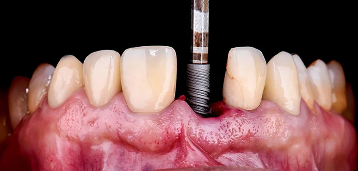
Free-hand surgery offers a cost-effective option. The clinician uses diagnostic information to open the flap and place the implant. This method uses intraoral periapical and panoramic radiographs to evaluate the alveolar bone width and other anatomy to place the implant. Free-hand surgery also uses the adjacent teeth as a guide, as well as using probes, calipers, etc., to determine the height and thickness of the ridge.
The benefits of free-hand dental implant surgery include:
FREE TO DOWNLOAD – SUCCESS GUIDES
DOWNLOAD A GUIDE
Free-hand Dental Implant Surgery Disadvantages
Free-hand implant placement offers several benefits because this method couples diagnostic data with onsite visualization. However, there are some drawbacks to this method, and they include:
The free-hand implant surgery method has been widely used over the years. With the invention of software and digital applications, a more accurate method for placing implants has emerged in the form of dental implant-guided surgery.
Guided Dental Surgery
There is no question that guided surgery workflows offer the highest level of control and accuracy.
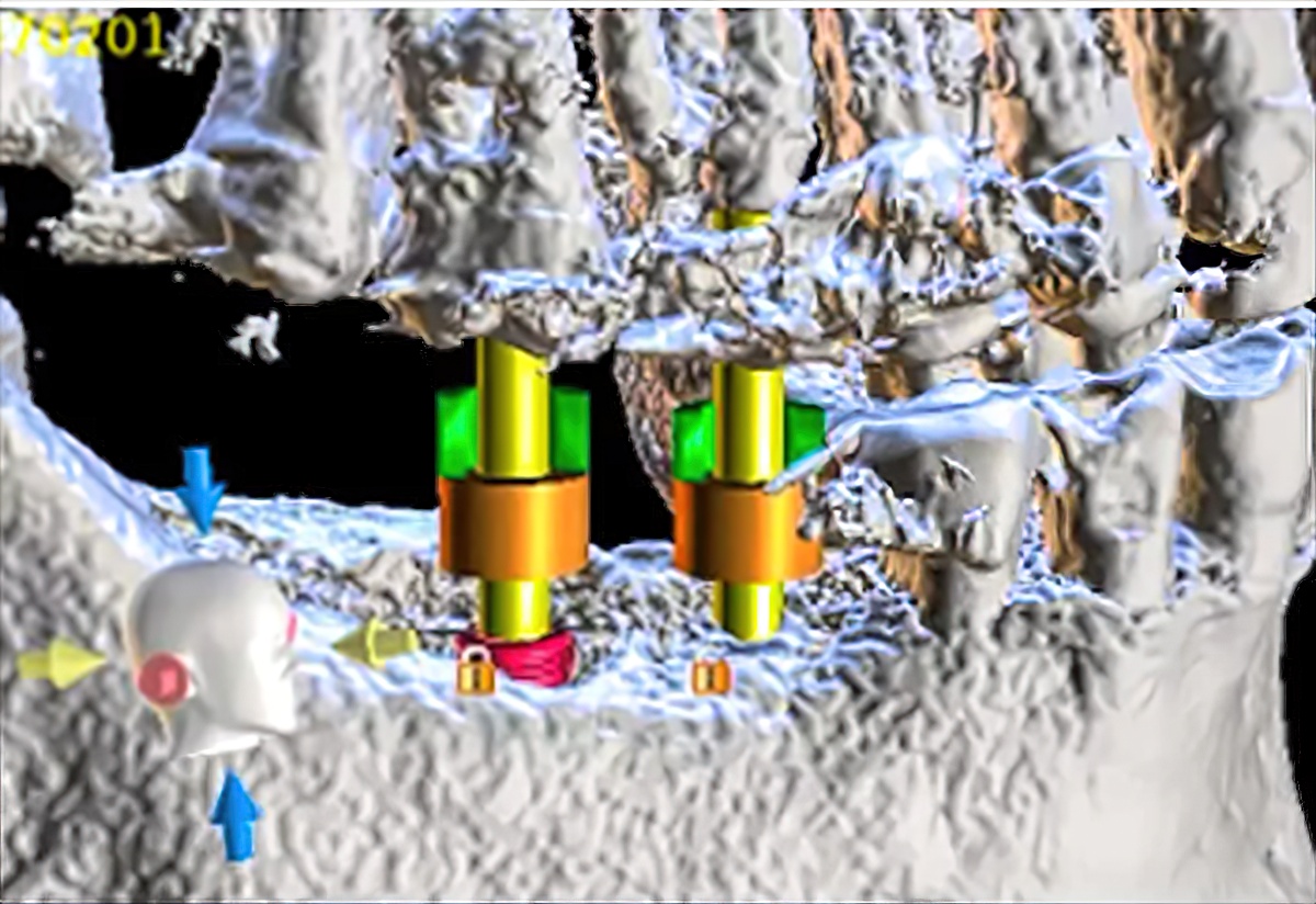
This method uses a digital surgery guide that helps the dental surgeon experience a safer, faster, more accurate, and in most cases, lower-cost surgical procedure. The software used in guided surgery provides a three-dimensional view of future implant placement. The procedure involves the patient having a cone-beam computed tomography (CBCT) scan. This data is then merged with surgical guide design software. This allows the clinician to see the bone density, nerves, and tooth roots of neighboring teeth. The ability to add this level of data to implant therapy cases makes the implant process much more reliable. The benefits of guided surgery include the following:
Once the CBCT scan is complete, a digital imaging and communications in medicine (DICOM) file is created. The DICOM file is sent in with a digital intraoral impression or a traditional impression, and a model is prepared.
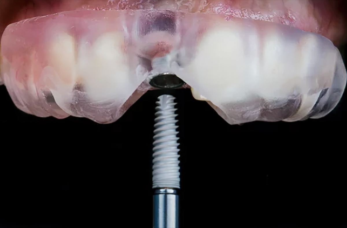
The software can then use the DICOM file and patient data to provide both two-dimensional and three-dimensional outputs. The data reveals important anatomical areas such as the maxillary sinus, submandibular fossa, and others.
Guided Dental Surgery Disadvantages
While guided dental surgery appears to offer many benefits over free-hand implant placement, there are some disadvantages to this workflow. For example, the software costs and tooling necessary to use this type of technology may be a drawback, and there is a learning curve.
Other disadvantages include:
Surgical Guide
The surgical guide is created specific to the patient’s situation. Factors such as whether the patient is completely edentulous or still has some teeth and the number of implants that will be placed will determine the type of guide produced.
Burbank Dental Lab fabricates surgical guides with round titanium sleeves embedded where the implants will be placed. These titanium sleeves provide the proper buccal-lingual location, mesial-distal location, angulation, and depth of the implants to be placed. They are unique to the type of surgical system being used.
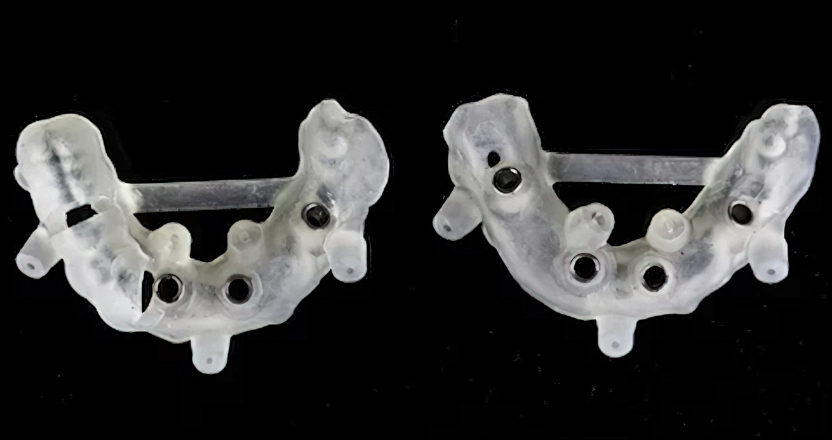
There are several different approaches to creating guided surgery workflows. Burbank Dental Lab uses the best digital printers on the market to be able to fabricate surgical appliances for these workflows. If you design your own guides or work with a third party, all that is needed is an .stl file of the design. Burbank Dental Lab will take the .stl and produce the appropriate guide.
Burbank Dental Lab has had great success working with Blue Sky Bio software. This is an open system that will allow the clinician to use any CBCT/DICOM file along with an .stl file of the arch to plan and design a surgical guide. Burbank Dental Lab’s guided surgery appliances offer the following benefits:
It is evident that while free-hand implant placement is done with success in many cases, guided surgery offers a high degree of accuracy. Both methods offer advantages and disadvantages. The key is to decipher if there are any chances for error and if so, guided surgery may be the best option. Call or chat with a team member today to learn more about Burbank Dental Lab’s implant services.
3 STEPS TO
PHOTOGRAMMETRY SCANNING
WE HELP YOU GET IT RIGHT
EVERY TIME
STEP 1
Burbank Dental Lab implant scan specialist provides the special scan bodies for insertion in the patient’s newly placed implant sites.
STEP 2
Scan Bodies are removed and special healing abutments are placed on the implant sites. Soft tissue can now be scanned with an intra-oral scanner.
STEP 3
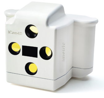
The data from both scans are then sent to the laboratory. The ICam4d data and soft tissue scans are aligned to become a high-precision dental model.
