



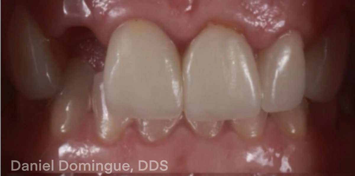
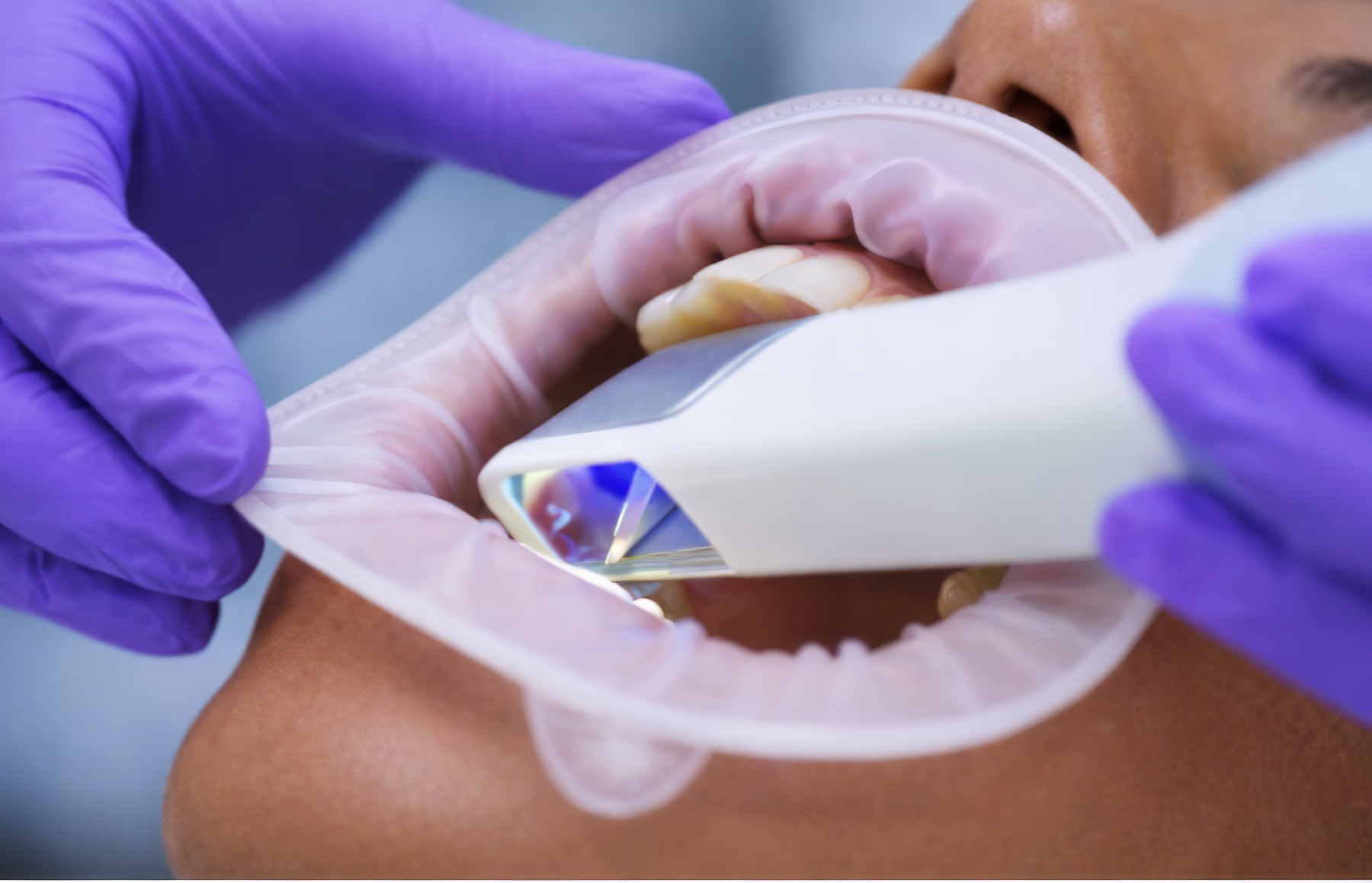

If the margin cannot be seen clinically, it will not be captured digitally.
A Clinical Guide to Achieve Accurate Crown Margin Capture for Dentists
Accurate margin capture is one of the most important factors affecting the fit and longevity of a dental restoration. Whether the restoration is a single crown, a veneer, or an implant restoration, the dental laboratory must clearly identify the preparation finish line to design a restoration that seats properly and seals the margin.
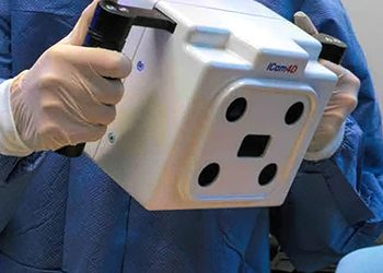
With the widespread adoption of intraoral scanners (IOS), dentists now capture digital impressions rather than traditional PVS impressions. Digital impressions offer many advantages, including improved workflow efficiency, faster communication between the lab and the dentist, and the ability to review scans immediately.
However, margin capture still depends on proper tissue management, preparation design, and scanning technique.
Dental labs review thousands of digital scans every year. One of the most common issues encountered during CAD design is incomplete or unclear margin data. When the finish line is not clearly captured, the laboratory must either request a new scan or estimate the margin location, which can affect the fit of the restoration.
Accurate digital margin capture typically depends on three clinical factors:
This guide explains how margin capture works in digital dentistry, why margin scans sometimes fail, and how dentists can improve margin accuracy when scanning with an intraoral scanner.
What is “margin capture” in digital dentistry?
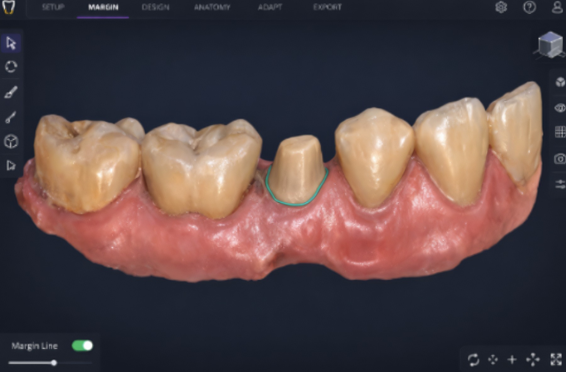
Margin capture refers to a digital scanner’s ability to accurately record the finish line of a tooth preparation. The finish line is the boundary where the restoration meets the prepared tooth surface.
For a dental laboratory to design a restoration correctly, the margin must be clearly visible around the entire circumference of the preparation.
Unlike traditional impressions, where the margin is captured within impression material, digital scanners rely on optical data. The scanner must be able to directly visualize the margin in order to record it accurately.
When margins are not clearly captured, several problems may occur:
Why does margin accuracy determine restoration fit?
Margin accuracy directly affects how well a restoration seats and seals at the tooth interface. During the digital workflow, dental laboratories must identify the preparation margin inside CAD software before designing the restoration.
This process is called margin detection.
Technicians examine the digital scan data and trace the finish line around the preparation. The CAD software then uses that margin line to generate the restoration design.
If the margin is unclear in the scan data, several problems can occur:
Digital workflows depend on precise scan data. If the finish line cannot be clearly visualized, the software cannot compensate for missing information.
For this reason, complete margin visibility is one of the most important factors affecting restoration accuracy in digital dentistry.
Are Digital Impressions More Accurate Than Traditional Impressions for Crown Margins?
Digital impressions and traditional impressions can both produce highly accurate crown margins when the preparation finish line is clearly exposed.
Traditional impressions rely on impression material flowing into the gingival sulcus to capture the margin. Digital impressions rely on optical imaging, which means the scanner must be able to directly visualize the margin.
In both workflows, proper gingival retraction and moisture control are essential for reliable margin capture.
The PMMA provisional isn’t a placeholder — it’s the working blueprint from which the final restoration is built.
Digital Margin Capture Depends on Visibility
With intraoral scanners, margin capture depends entirely on optical access. The scanner cannot detect margins hidden beneath gingival tissue or obscured by fluids.
For this reason, dentists using digital impressions must focus on several clinical factors before scanning:
When margins are clearly exposed and scanned carefully, digital impressions can produce restorations with excellent marginal accuracy.
Digital workflows depend on precise scan data — if the finish line can’t be visualized, the software can’t compensate for missing information.


The objective is simple: the entire margin must be clearly visible before scanning.
Why Dental Labs Prefer Clear Digital Margins
From a dental lab perspective, digital impressions often provide excellent detail when margins are clearly captured.
Technicians can zoom into scan data, rotate the model, and precisely trace the finish line during CAD design. This can allow for very precise restoration fabrication.
However, when the margin is unclear in the scan data, the lab cannot compensate for missing information. In these situations, the dental lab may need to request a new scan before fabrication can proceed.
Digital workflows are most predictable when dentists verify that the entire margin circumference is clearly visible in the digital scan before submitting the case.
Common Reasons Intraoral Scanner Margin Scans Fail
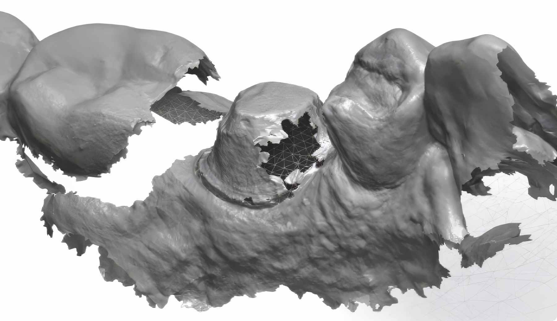
Digital margin scans most commonly fail due to soft tissue interference or contamination around the preparation. From a lab perspective, these issues are often immediately visible in the scan data.
Common causes include:
When dental labs review scans with these issues, lab technicians may not be able to identify the true margin location.
Digital Scanner Tracking and Stitching Errors
Intraoral scanners create a 3D model by stitching together thousands of images captured during scanning.
If scanning movement is inconsistent or the scanner loses tracking, stitching errors may occur. These errors can distort surface data near the margin and make the finish line difficult to interpret in CAD software.
Scanning slowly and maintaining continuous scanner tracking helps minimize stitching artifacts and improve margin accuracy.
Tissue Management Techniques for Margin Exposure
Even in digital workflows, traditional tissue management remains critical. If the margin cannot be seen clinically, it will not be captured digitally.
Several techniques are commonly used to expose margins before scanning.
Digital Cord Technique
The dual cord technique involves placing two retraction cords.
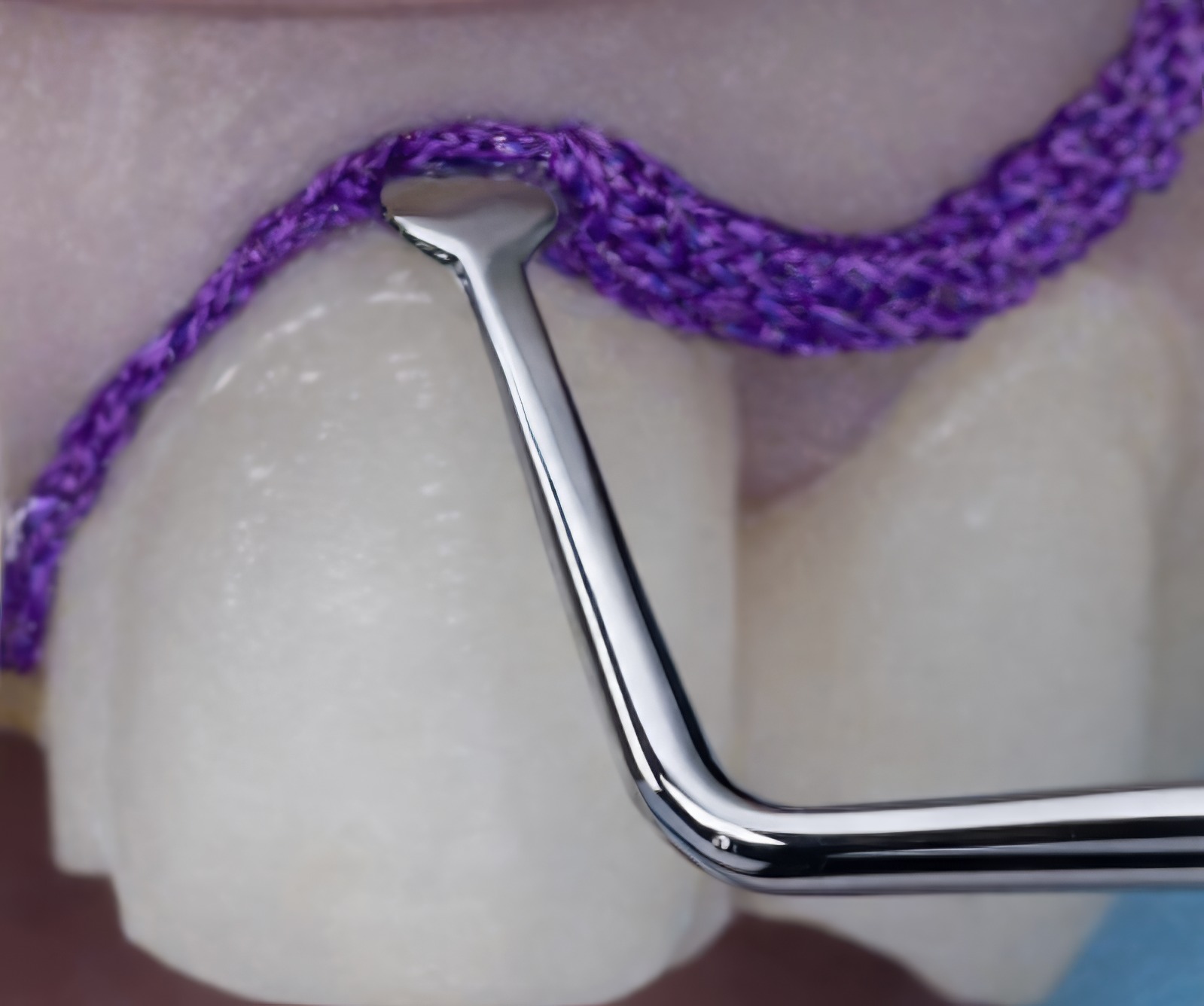
The upper cord is removed before scanning, while the lower cord remains in place to maintain tissue displacement.
This technique often provides the most reliable margin exposure.
Single Cord Technique
A single retraction cord can provide adequate tissue displacement for shallow margins but may offer less control than the dual-cord technique.
Retraction Pastes
Retraction pastes can help control bleeding and gently displace gingival tissue. They may be useful when traditional cord placement is difficult.
Laser or Electrosurgical Tissue Management
Soft-tissue lasers or electrosurgical units may be used to remove tissue that obstructs margin visibility. These techniques can be helpful when margins extend slightly subgingivally.
Regardless of the technique used, the objective is simple: the entire margin must be clearly visible before scanning.
Digital dentistry works best when dentists and laboratories collaborate to produce high-quality scan data.
Preparation, Design, and Margin Placement
Preparation design significantly influences digital margin detection.
Margins that are clearly defined and easily accessible are more likely to be captured accurately.
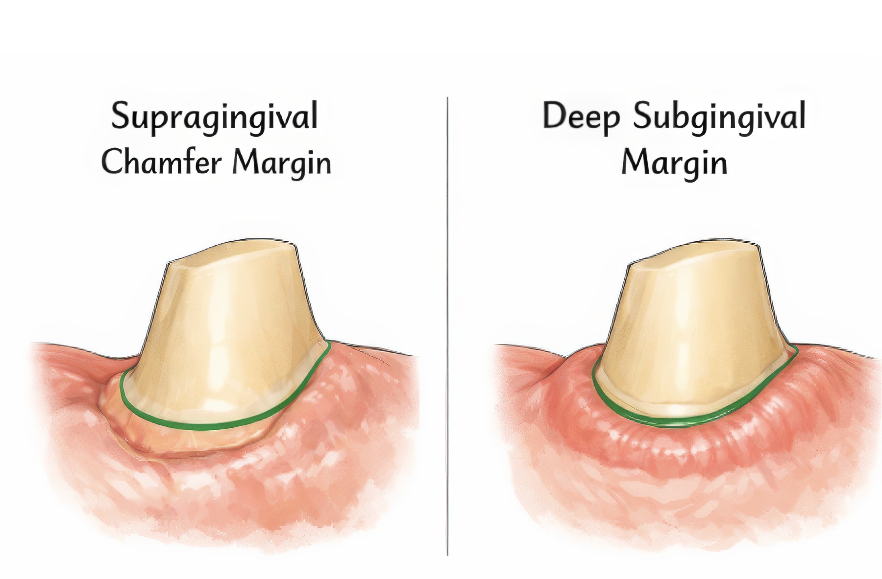
Supragingival margins
Supragingival margins are the easiest to scan because the finish line is fully visible. They provide the most predictable digital margin capture.
Equigingival margins
Equigingival margins may be captured successfully when tissue management is adequate.
Deep subgingival margins
Deep subgingival margins are the most difficult to capture digitally because the scanner cannot visualize margins covered by tissue.
Margin geometry
Margin shape also influences digital detection. Chamfer margins often produce clear edge definition for scanners. Shoulder margins can also scan well when clearly exposed.
Margins with irregular geometry or poorly defined finish lines may be more difficult for CAD software to interpret.
Recommended Scan Path for Crown Preparations
Scan path strategy plays an important role in the accuracy of digital impressions.
Many clinicians find that following a consistent scan path improves scanner tracking and reduces stitching errors.
A commonly recommended scan path includes:
Maintaining continuous scanner movement helps the software build a stable digital model.
Intraoral Scanner Technique for Margin Capture
The operator-scanning technique also affects the quality of margin capture. Even when the margin is clinically visible, an improper scanning technique can prevent the scanner from recording it.
Several techniques can improve margin accuracy.
Verify Margin Capture in the Digital Model
After completing the scan, clinicians should inspect the digital model before sending the case to the lab.
Rotate the model and zoom into the preparation to confirm that the entire finish line is clearly visible.
Many dental labs report that margin issues could be avoided if clinicians carefully reviewed the scan data before submission.
What Dental Labs Need from Digital Scans
Dental labs rely entirely on scan data to design restorations. Technicians cannot physically examine the preparation and must depend on the digital model.
When margin clarity is poor, several challenges may occur:
In some cases, laboratories must request a new scan before proceeding with fabrication.
Clear margin scans allow technicians to confidently identify the finish line and design restorations with proper seating and marginal integrity.
Digital dentistry works best when dentists and dental labs collaborate to produce high-quality scan data.
Clinical Checklist for Better IOS Margin Scans
Before scanning a crown preparation, dentists can confirm several key conditions to improve margin capture.
Before scanning, confirm:scan data.
Taking a few moments to verify these factors can significantly reduce the need for restoration remakes.
The Future of Digital Margin Detection
Digital dentistry continues to evolve rapidly. Modern intraoral scanners are improving in resolution and image processing capabilities.
Several developments may improve margin detection in the future:
Although these technologies will improve digital workflows, accurate margin capture will continue to depend on good clinical technique and proper tissue management.
The best digital margin isn’t fixed in the lab — it starts with what the scanner was able to see.
FREE TO DOWNLOAD – SUCCESS GUIDES



DOWNLOAD A GUIDE
FAQ


 Previous Post: