There is no question that digital dentistry has changed dentistry for the better. To provide the best patient experience, a move to digitizing workflows is one of the most important investments a dental office can make. The intraoral scanner is by far the easiest and best place to start making the transition from traditional dental workflows to digital dental workflows.
The Benefits of Intraoral Scanners
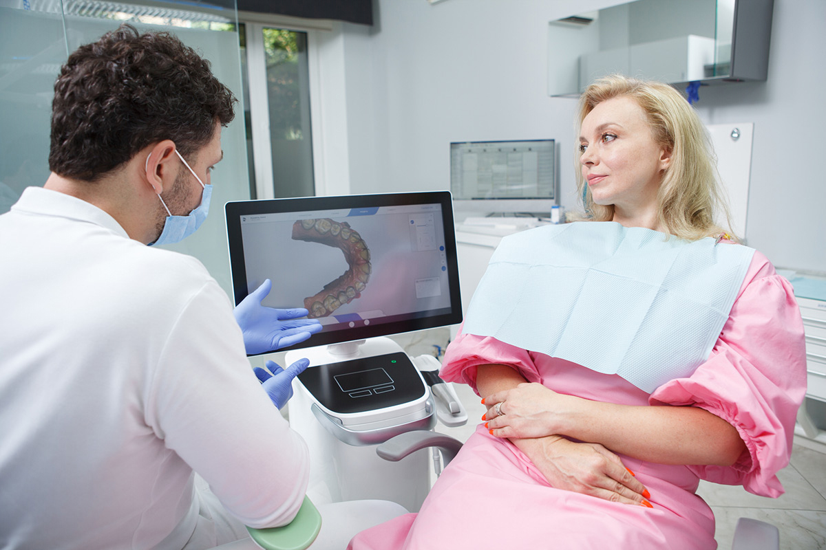
Digital impressions offer advantages throughout the entire dental process. The intraoral scanning process benefits the dental office, the patient, and the laboratory.
Some of these benefits are:
Things To Consider When Choosing An Intraoral Scanner
Making the decision to move to digital technologies is an excellent option for a dental practice. An intraoral scanner is an excellent way to get started in digital workflows. It can be daunting to decide which type of intraoral scanner to choose, but here are some guidelines to help make the best decision:
1. Accuracy of the intraoral scanner.
This may be by far the most important consideration. The quality of the scan is directly related to the quality of the final restoration.
The scanner must capture details accurately to ensure excellent restorative outcomes.
2. Ease of use.
The easier an intraoral scanner is to use, the more seamless the integration into current practice workflows will be. The software should be easy to work with and easy for the entire staff to manage.
3. Software considerations.
The software is essential to the usability of the scanner. This is often evaluated based on the speed of the scan. There should be no lag time between the scan and the visual output of the images on the computer screen.
4. Intraoral scanner design.
Busy dental practices should consider the ergonomics of the scanning wand. It should be easy to hold and maneuver within the patient’s mouth.
The wand should also be lightweight to help eliminate the possibility of fatigue. The scanning wand should also be designed to ensure the patient has a pleasant experience.
5. Support and training.
There will be a learning curve. It is important to choose a company that has an excellent training program and an ongoing support system as you become more familiar with the scanner. In addition, ask about the equipment warranty and technical support.
6. Financial considerations.
Many of the intraoral scanners have similar price options. There are other costs, however, that should be evaluated before making a decision. These include the following:
- Will I need any additional equipment?
- Do I need to pay for technical support?
- Are there any recurring costs, such as software subscriptions?
With a little research, these guidelines can help provide the best intraoral scanner system that suits the unique needs of any dental practice.

Tips on Getting the Most Out of Your Intraoral Scanner
Intraoral scanners benefit the dental practice, the patient, and the lab. They can be used for providing scans necessary for the fabrication of crowns, bridges, partials, nightguards, and implants. They do, however, have a learning curve. Here are some tips on navigating through the curve:
1. Practice
It will take some time to adjust to using the scanner. The best way to get experience is to use the device and practice. Take gradual steps that will allow you to make adjustments as needed.
The more you use it, the more it will become second nature.
2. Make sure to scan in an extremely dry field.
Moisture will seriously impede the accuracy of digital impressions. Use suction and compressed air just before taking the scans.
3. Make sure the scanning wand is dry.
Make sure the scanning wand is dry and clean before scanning.
4. Retraction of the tissue around the preparation is extremely important.
An intraoral scanner can only pick up what is visible. It is advised to use two-cord retraction to get clear scans.
5. Take a pre-op scan.
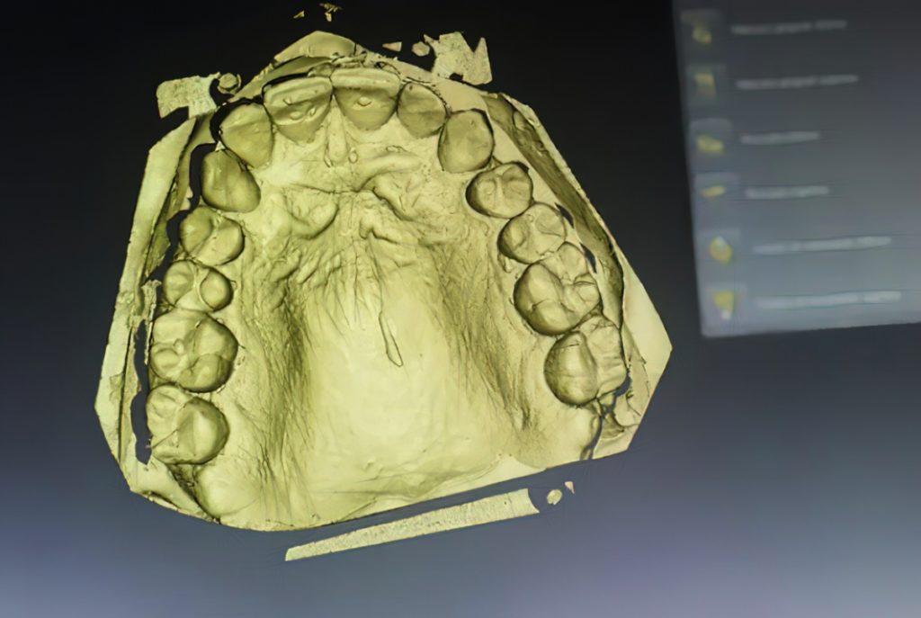
This can help the lab during the designing of the new restoration. This way, the lab can recreate the contour and shape of the natural tooth as appropriate. In addition, provide the following scans:
- The arch with the prepared tooth
- The opposing arch
- The bite or the buccal view in maximum intercuspation
6. Scanning process requires a slow, steady approach.
The scanning process itself requires a slow, steady approach. It is important to stay in each area until the required anatomy is recorded. If you find you are seeing holes or excess material in the final image simply slow down. Most issues on digital scans are caused by not staying in an area long enough to capture the data.
7. Rotate the scanning wand to avoid missing gingiva.
A fairly common issue for new users is missing gingiva. This can be remedied simply by adequately rotating the scanning wand until 4-5mm of the tissue is captured.
8. Analyze scans.
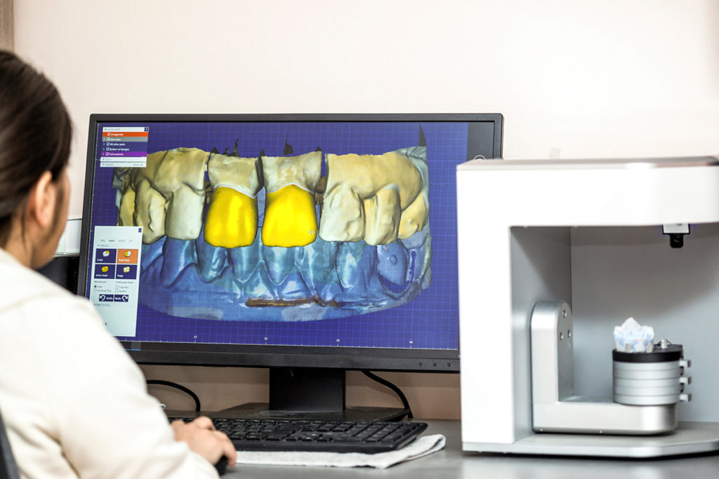
Analyze the scans before sending them to the lab.
Burbank Dental Lab accepts intraoral scan files from any intraoral scanning system, including the following:
- Itero
- Trios
- 3M
- Medit
- Cerec
- Carestream
- Straumann Cares
If you are using a system not mentioned above, Burbank Dental Lab can receive any STL file. Simply send it to our digital team at digital@burbankdental.com.
Making the transition from traditional impression methods to digital impressions can seem daunting. However, the technology and support are available to make this move seamless. Adopting intraoral scanning technologies in a dental practice makes for improved accuracy, patient experience, perceived value, communication, and case acceptance.
At Burbank Dental Lab, our digital team can answer all your questions regarding going digital.

