The Face-Bow That Can Make A Difference

mm Written by Andrew Sedler

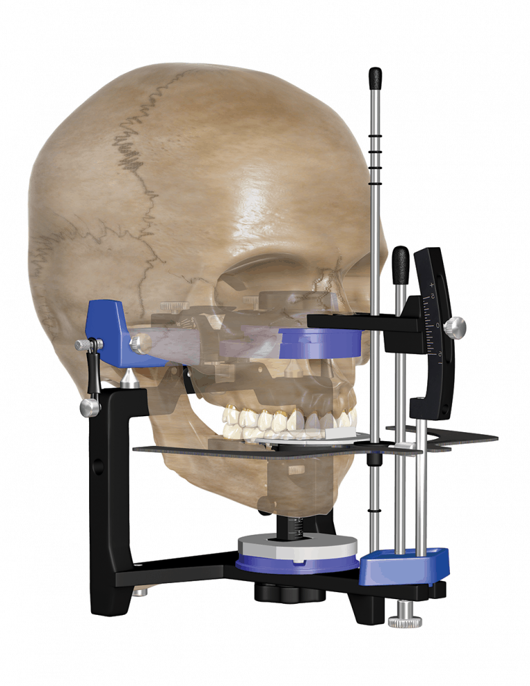
Burbank Dental Kois Dento-Facial Analyzer

The Kois Dento-Facial Analyzer

Dental labs like Burbank Dental Lab rely on the records that the clinician collects and sends to us. Our technicians can replicate the function and esthetics of natural dentition, but we can be limited by the information communicated to us through the patient records.

Whether you are creating a beautiful smile make-over with IPS e.max, or restoring a patient’s whole mouth, you will want to communicate essential information to the lab. There are four essential elements that the lab requires to deliver the most predictable restorations:

  1. Photos: full-face smile, smile close-up, and lip at rest
  2. Pre-Op models
  3. Bite records
  4. A Face-Bow record

This article will focus on the face-bow record, specifically, the Kois Dento-Facial Analyzer. The advantage of a face-bow mounting is to place the dentition in a 3-D relationship to the articulator’s condyle. However, too often the esthetics of the smile line is lost. One advantage of the Kois Dento-Facial Analyzer is that it facilitates both functional and esthetic objectives.

First let me say, use of a face-bow is not required for every case. There are, however, three scenarios that need the additional information that a face-bow provides.

  1. The distal terminal abutment, especially a bridge.
  2. Full arch rehabilitation.
  3. Smile makeover cases.

Note: Full arch impressions are assumed for the above cases and any use of a face-bow.

Burbank Dental Lab

SPECIAL OFFER

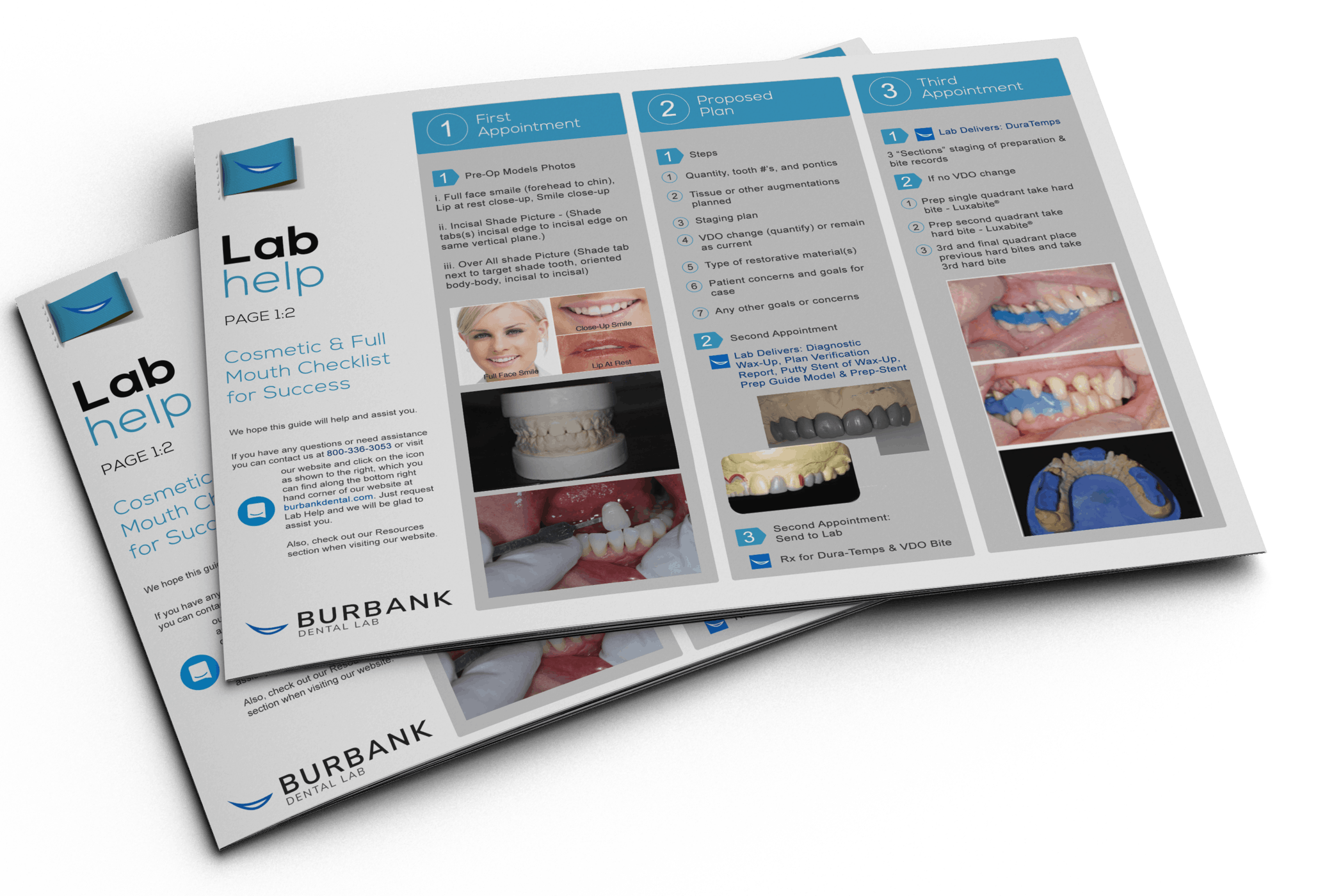
Cosmetic & Full Mouth Checklist for Success by Burbank Dental Lab

FREE Case Planning Guide & Workflow Chart

OFFER GOOD THROUGH APRIL 16, 2018
Day
Hour
Minute
Second

FREE Case Planning Guide and Workflow Chart

Burbank Dental Lab

What Makes the Kois Bow Different?


Simplified bow design & procedures

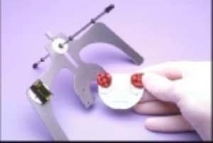
The Kois Dento-Facial Analyzer is an elegantly simple instrument. Unlike other face-bows, there is no ear bow, movable joints, or tightening screws.  “Kois has developed a unique face-bow transfer that corresponds to the facial midline and horizontal plane to the 100-mm distance of the conditional hinge position. This technique simplifies the process of the face-bow transfer and ensures the occlusal plane of the teeth is evaluated and fabricated correctly.” Ref. 1 (Figures 1-3)

Kois Bow instructions
Burbank Dental Lab, Kois Bow instructions
Burbank Dental Lab - Kois Bow instructions

The basic clinical steps to using the Kois Dento-Facial Analyzer are as follows:

  1. With the posterior portion of bow out of occlusion, set the incisal edge of maxillary incisors to the ledge on Index Tray. The incisal ledge is designed to position the incisors based on an average 100mm distance from the condyle and the central incisor edge.
  2. Align Vertical Indicator Rod to patient’s facial midline to register the dental midline of the teeth.
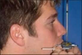
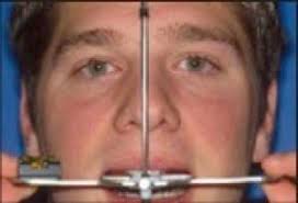
  3. While maintaining incisal contact with the Index Tray and vertical rod alignment to the facial midline, rotate Analyzer Bow up in the posterior until the lateral wings are level to the horizon. There is no need to take a stick-bite. The Kois Dento-Facial Analyzer captures the horizontal smile line, but it also places it into a relationship to the condyle. (Figures 4 & 5)
Kois Dento-Facial Analyzer
Burbank Dental Lab Kois Bow procedures
  • With the patient standing upright, confirm that the bow maintains proper position.
  • The central incisors are in contact with the incisal ledge on the Index Tray.
  • Ensure the horizontal plane of the bow is parallel to the horizon. (This can be assisted if you have patient stand next to a door jam and reference the vertical line of the door frame, keeping the bow perpendicular to this reference.)
  • Check the vertical rod in relation to the facial midline.
  • Have the lateral wings extend parallel to the horizon.

You have now captured the steepness and tilts of the occlusal plane in the registration material on the horizontally aligned Index Tray. Remove Index Tray from Analyzer Bow and send to the lab for mounting of study casts. This disposable Index Tray now becomes a permanent bite fork registration record.(Figure 6)

Burbank Dental Lab Kois Bow instructions

Error-Free lab procedure to transfer face-bow recordings to the articulator

Eliminating potential error in the transfer of the face-bow orientation is one of the strengths of the Kois bow.

The lab procedure is four simple steps.

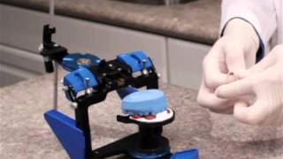
  1. Place the magnetic adjustable mounting platform onto the Panadent articulator’s lower mounting plate, then snap the face-bow transfer index tray on the adjustable platform. (Figure 7)
  2. Set the mounting platform to zero.
  3. Affix the maxillary model onto the index.
  4. Set anterior pin to zero and mount upper model to the articulator.
Burbank Dental Lab, Kois Bow transfer procedure

You are not sacrificing accuracy for ease of use.

  1. The evidence supports the accuracy of the Kois Face-Bow. “The Kois Dento-Facial Analyzer articulates the maxillary cast in a position that is not statistically different to the Pana-Mount face-bow when comparing the incisal edge position and occlusal plane angle relative to Frankfort Horizontal.” Ref. 2
  2. "The Kois Dento-Facial Analyzer is a quick, simple, and predictable way to mount the maxillary model to ensure functional and esthetic predictability." Ref. 3
  3. There is no risk of your face-bow transfer jig shifting in transporting it to the lab. With the Kois system, you do not send a jig, you send the Index Tray only.
  4. The lab transfer process is 100% predictable. There is no complicated transfer apparatus. The lab has a straightforward procedure of snapping the Index Tray onto the magnetic mounting table, affixing the model to the Index Tray and mounting.


Why you should adopt it into your tool belt.

  1. Simplicity - both the clinical and lab procedures are simple and reduce errors.
  2. Face-bow records taken with the Kois Dento-Facial Analyzer are duplicatable if there is a need to retake a second face-bow record.
  3. There are no movable parts in the transfer of the Index Tray.
  4. Combines the function of a face-bow transfer and a stick-bite in the same record.
  5. The Kois Bow establishes the midline of the smile.

In the final analysis, using the Kois Dento-Facial Analyzer in conjunction with Burbank Dental Lab will enhance your predictability and level of occlusal accuracy, while at the same time increasing your esthetic results.

For information about purchasing the Kois DentoFacial Analyzer contact Panadent Corp. http://www.panadent.com, or Burbank Dental Lab.

References:

  1. Kois Dento-Facial Analyzer System, Panadent Corp., Grand Terrace, CA. http://www.panadent.com
  2. Lux, Laura H., "A Comparison of Two Methods to Articulate a Maxillary Cast with Lateral Cephalometry" (2014).Master's Theses (2009 -). Paper 252. http://epublications.marquette.edu/theses_open/252
  3. KOIS DENTOFACIAL ANALYZER: HOW TO VIDEO, By Lee Ann Brady on 12.15.14 https://leeannbrady.com/occlusion-tmd/kois-dentofacial-analyzer-how-to-video

Did you like this article?

Let us know by giving us a clap!

Top 10 Most Clapped Articles

Resource Center

Products

Slide 1

Fabrication
of the future
is here!

Burbank Dental Lab has three new state-of-the-art Carbon M2 printers. We are very excited about the options that these cutting-edge printers will allow us to offer our dental clients. Here are some of the advantages that these printers will begin to deliver to you and your dental practice.

Our New
State-Of-The-Art
3d Printers

Carbon offers a highly dependable 3D manufacturing solution for many dental applications with its breakthrough Digital Light Synthesis™ technology, enabled by a wide range of dental materials.