


Guided surgery is one of the most discussed issues in dentistry. At Burbank Dental Lab, questions related to guided surgery are the number one area that we assist our clients. Developments in this area are moving at light-speed; it can be easy to get lost in the details, especially when the details change continuously.
One of the primary steps in the workflow of guided surgery is the CT scanning procedure; a radiographic scanning guide is recommended for this procedure. As with most successful treatment plans, the foundation of a guided surgery plan often begins with a diagnostic wax-up. (Figure 1) Whether for a single tooth or a full arch edentulous case, the wax-up is the template for the scanning guide, the implant placement, as well as the final restoration(s).

Figure 1
Once the initial wax-up is complete, it is used to fabricate the scanning appliance for use during the CT scanning appointment. The purpose of the scanning appliance is to identify in the CT, where the proposed tooth placement is desired. This will influence the planning and ultimately the placement of the implants to accommodate the final prosthetics.
4 Styles of Scanning Appliances
- Acrylic with barium sulfate teeth.This scanning guide is made from clear acrylic. The teeth to be supported by the implants should be made with 10% barium sulfate acrylic. The barium sulfate will make the teeth radiopaque for easy identification in the CT image. (Figure 2)
.
Figure 2
.
- Acrylic guide with added gutta-percha channel.
.
.
This technique for a scanning appliance, which has been used for many years, is to create the acrylic guide but to add a gutta-percha channel to identify the proposed implant angulation. (Figure 3) This technique is acceptable; however, fabricating the whole tooth out of a barium sulfate mixture allows for a more clear replica of the proposed tooth in relation to the screw access hole during digital planning.
.Both options 1 and 2 are acceptable if you are going to do your surgical plan and guide design via an open design software like Blue Sky Plan. Blue Sky is an innovative open architecture system that is free to the user, charges only occur only when you convert to a .stl file for printing or milling of your final guided surgery appliance.
.Figure 3
.
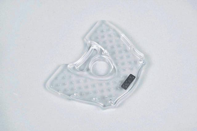
- SICAT plate scanning guide
.
This guide is used exclusively in the GALILEOS / SICAT ClassicGuide workflow. The SICAT scanning guide and subsequent ClassicGuide are particularly useful for edentulous cases. This guide uses the SICAT bite plate that is fused to an acrylic appliance which incorporates the tooth set-up. The advantage of this approach is that the SICAT bite plate has radiographic markers built in for added precision. Within this process, SICAT is sent the scanning guide, models and the DICOM file from the CT scan. SICAT does the plan according to the clinician's Rx and allows the doctor to make final approval before the fabrication of the ClassicGuide, which is only fabricated by the SICAT lab in Germany. (This workflow is only available to clinicians using a Sirona CT scanner.)(Figure 4 & 5)
Figure 4
.
Figure 5
- Don't use a scanning guide at all
The diagnostic wax-up has previously been limited to just that, a wax-up on a stone model. Now there are digital choices for this part of dental implant planning. Systems like CEREC and Blue Sky Plan allow you to digitally place teeth in the position of the desired restoration digitally. This option of "digitally waxing-up" cases is limited to partially edentulous cases that will have a tooth-borne guided surgery appliance..
a. SICAT no radiographic scan guide option
Via the GALILEOS / SICAT / CEREC and Cerec Guide 2 workflows it is possible to avoid the need for a scanning guide all-together. Scanning without the need for a scanning guide is accomplished in the OptiGuide and DIGITALGUIDE workflows, by adding the teeth in digitally in the planning phase. This is accomplished by using CEREC design software to add in the proposed addition of teeth that will be supported by the implants. In this process scan your patient with a Sirona 3D system and record the optical surface data. A radiographic template or other reference bodies are not necessary. (Figure 6)
.
Figure 6
.
b. Blue Sky Plan no radiographic scan guide optionWhen using Blue Sky Plan (BSP) software, it is also possible to avoid the need for a radiographic scanning appliance. You can add teeth from within the BSP software; this must be for a tooth-borne guide..There are limits on what type of case can be successfully planned without a physical mock-up.In speaking with Dr. Armen Mirzayan (Owner of Cad-Ray.com), he sets limits for not requiring a radiographic guide as follows: it is not required "...only if there are less than 5 teeth that are not heavily restored with radiopaque material. If all teeth have crowns, they will too much scatter, so you will need a scan appliance."


Overview
.There are a few advantages to using a mock-up directed radiographic scanning guide. Beginning with a wax-up or duplicate of a denture can be a useful tool that allows you to focus on an ideal restorative conclusion before the scanning appointment. When this wax-up/mock-up is used to create a radiographic guide, the ideal outcome is incorporated into your CT-based surgical plan and will produce a more predictable outcome. The more you plan at the start of the case, the less chance of running into surprises at the end.


